Comparing Apical and Basal Secretome Levels in Static vs. Apical Flow Conditions
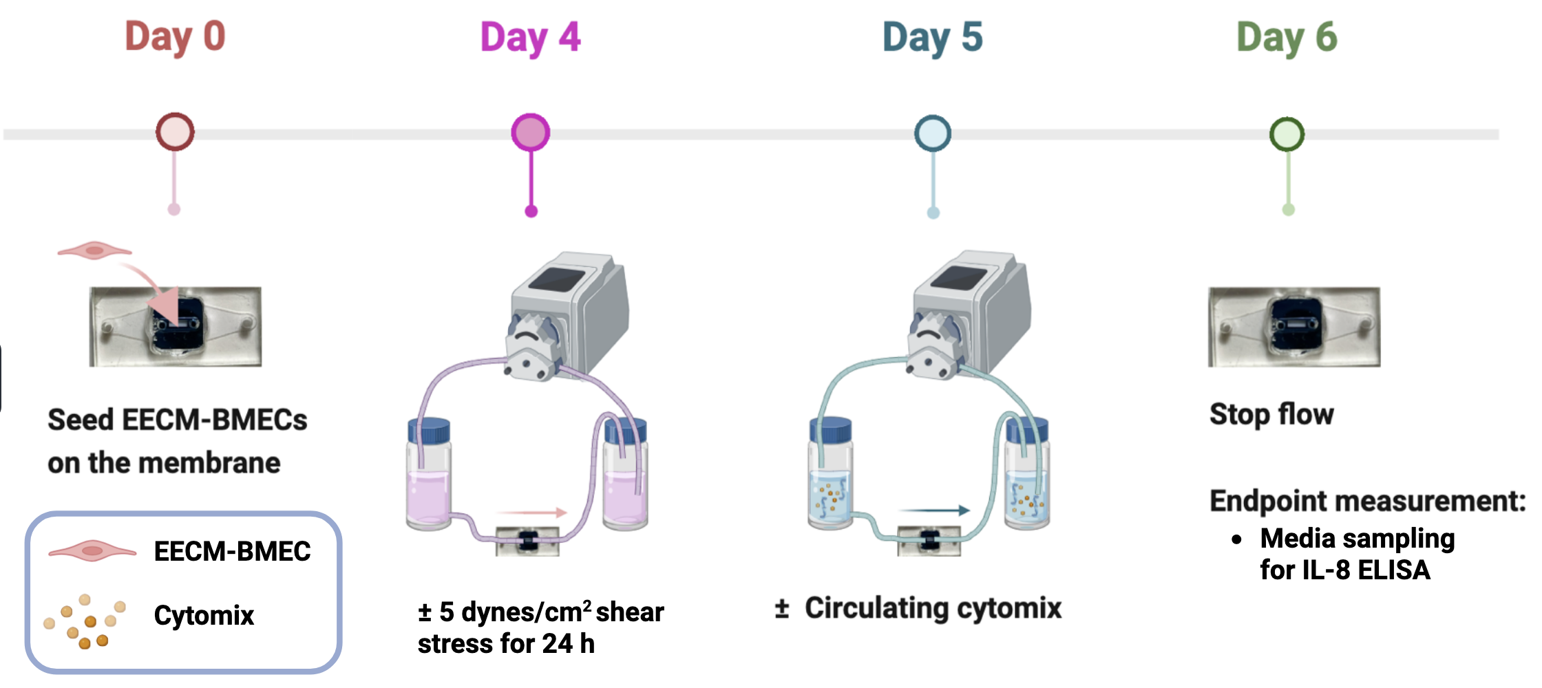
We’ve long speculated that adding fluid flow over our tissue models would reduce the concentration of pro-inflammatory factors secreted in response to inflammatory challenges, dampening the overall inflammatory state compared to static culture. We now have evidence that this is true and the results are important as benchmarks for our future studies. In response to reviews for Chloe Chen’s recent publication , Isabelle conducted an experiment to measure IL-8 secretion into the apical and basal sides of the µSiM under static and flow conditions ± cytomix (50 pg/mL of IL1-β, ΤΝF-α, ΙNF-γ). The culture system in these experiments was a simple monolayer of iPSC-derived BMECs. Isabelle used ELISA for the assessment of IL-8 secretion into the apical and basal compartments. The culture timeline is depicted below:
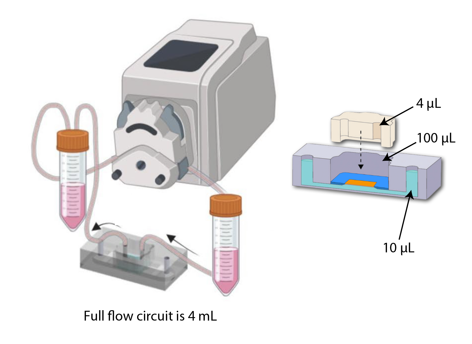
Here is the standard curve: 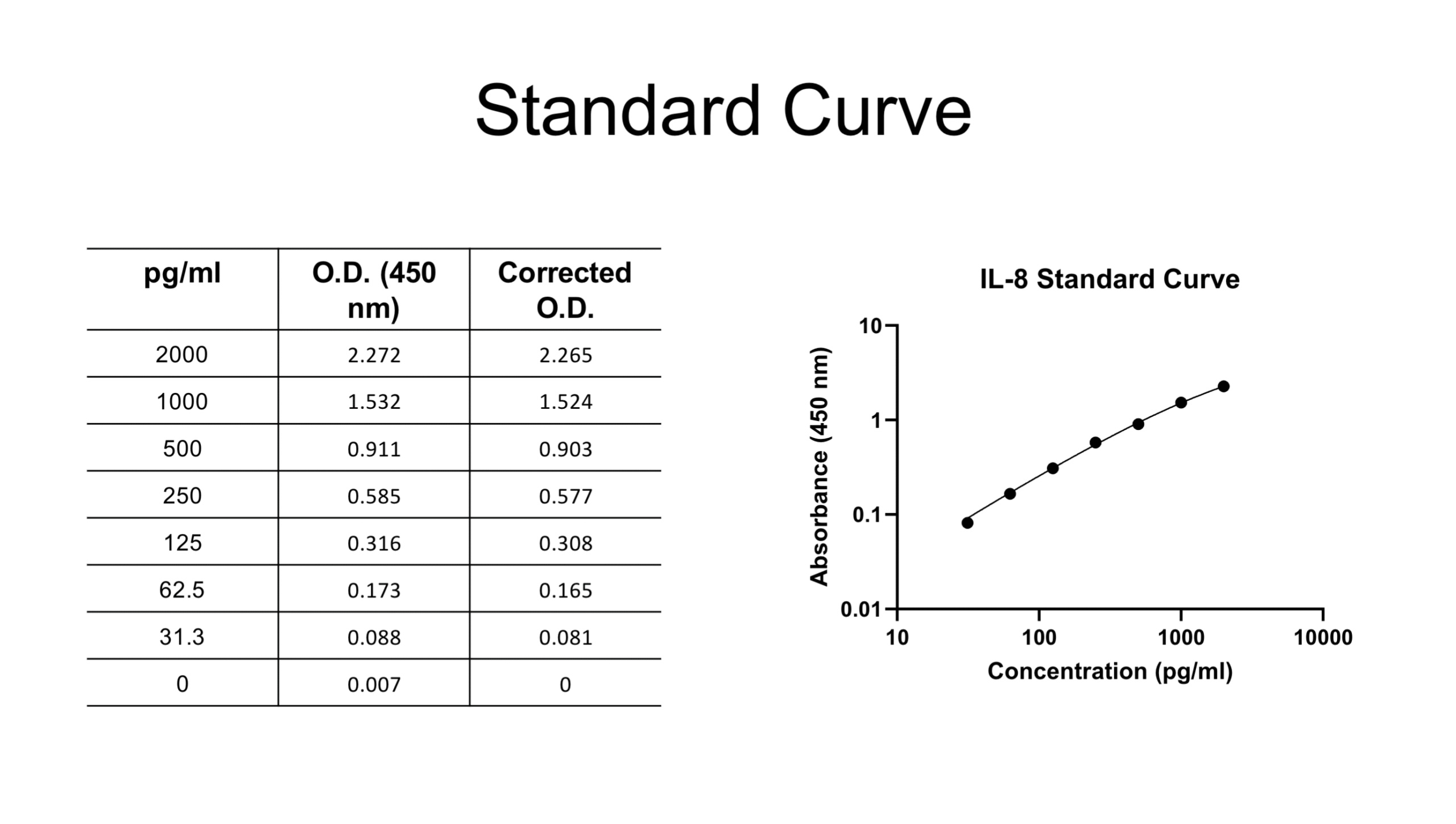 One concern we had going in was the ability to harvest enough IL-8 from the device to be detected by ELISA. The curve tells us that 30 pg/mL and higher are safe to interpret. We harvested IL-8 from the basal compartment using the ‘reverse pipette’ technique that we developed for permeability measurements. For this experiment, we added 200 µL of hECSR media to a pipette tip attached to one bottom channel port, acting as a reservoir. 200 µL of media, including the 10 µL bottom channel volume, was collected by reverse pipetting from the opposite port. For our concentration calculations, we accounted for this dilution by multiplying the assay output by 20. To collect media from the apical flow channel of static devices, the 50 µL media droplet on top of the insert was removed. Then, 50 µL fresh media was flushed through the flow channel and collected via the opposite flow port. To account for the dilution factor, we multiplied assay outputs by 25. For flow conditions, apical measurements were taken by withdrawing sample directly from the upstream reservoir. The volumes of the flow circuit and each of the device components will contribute to final concentrations.
 Results are shown graphically and in the a table below.
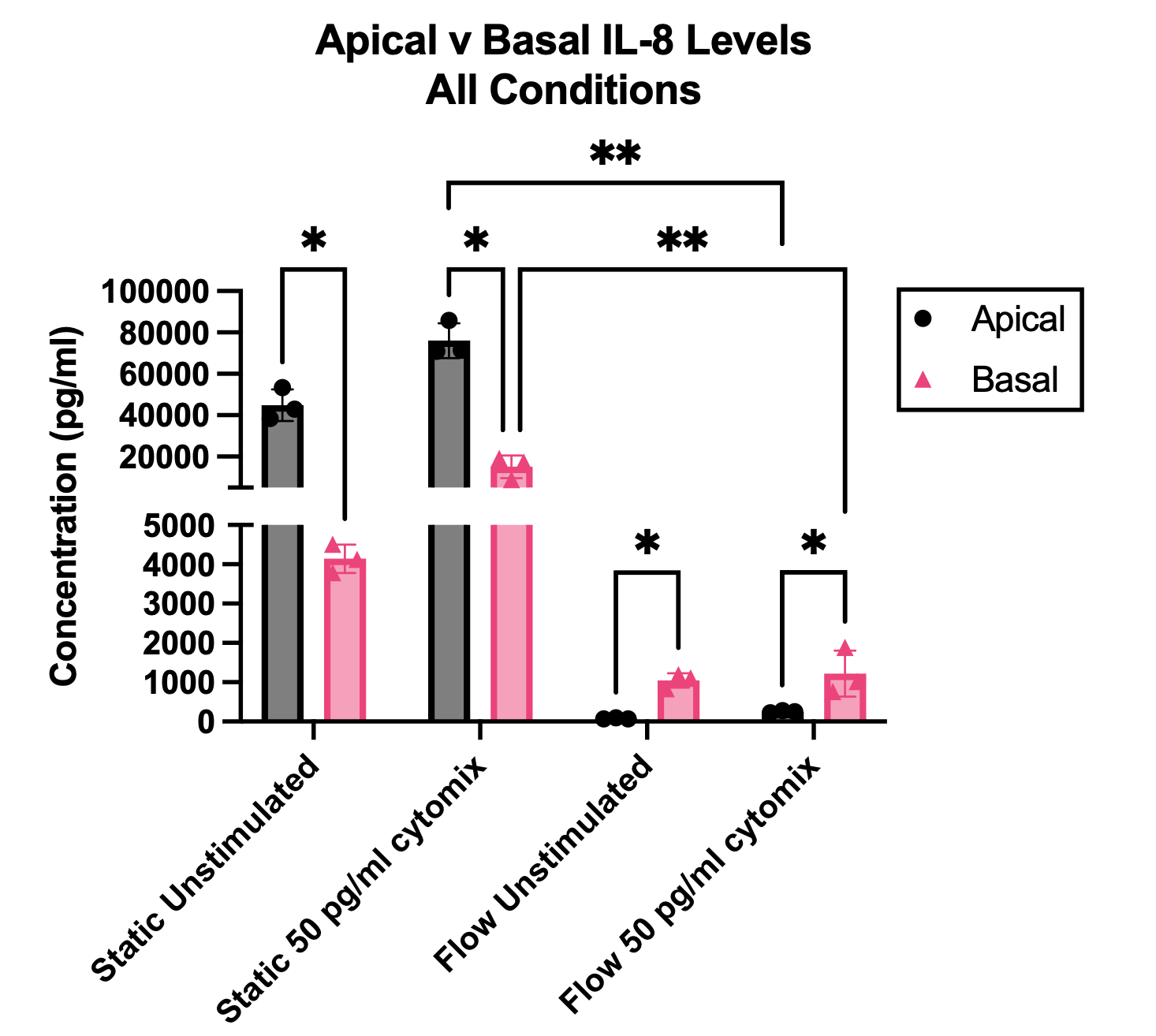
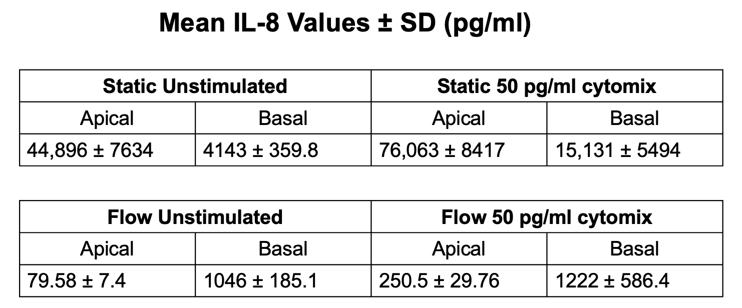
The first thing to note is that the static culture IL-8 concentrations are orders of magnitude higher than the flow based cultures in both the apical and basal samples. Additionally, the static culture IL-8 levels are orders of magnitude higher than the applied inflammatory challenge (50 pg/mL each of IL1-β, ΤΝF-α, ΙNF-γ))! Under flow, the IL-8 levels in both apical and basal compartments are comparable to the applied stimulation.
Thus in static culture, a supraphysiological secretome dominates the culture. In flow things are tuned to a more physiological level thanks to clearance and dilution as happens in the body. However, our choices of volumes, cell numbers, etc. are all arbitrary. So how do we know that we’ve got these balances right? We don’t. More thinking is required.
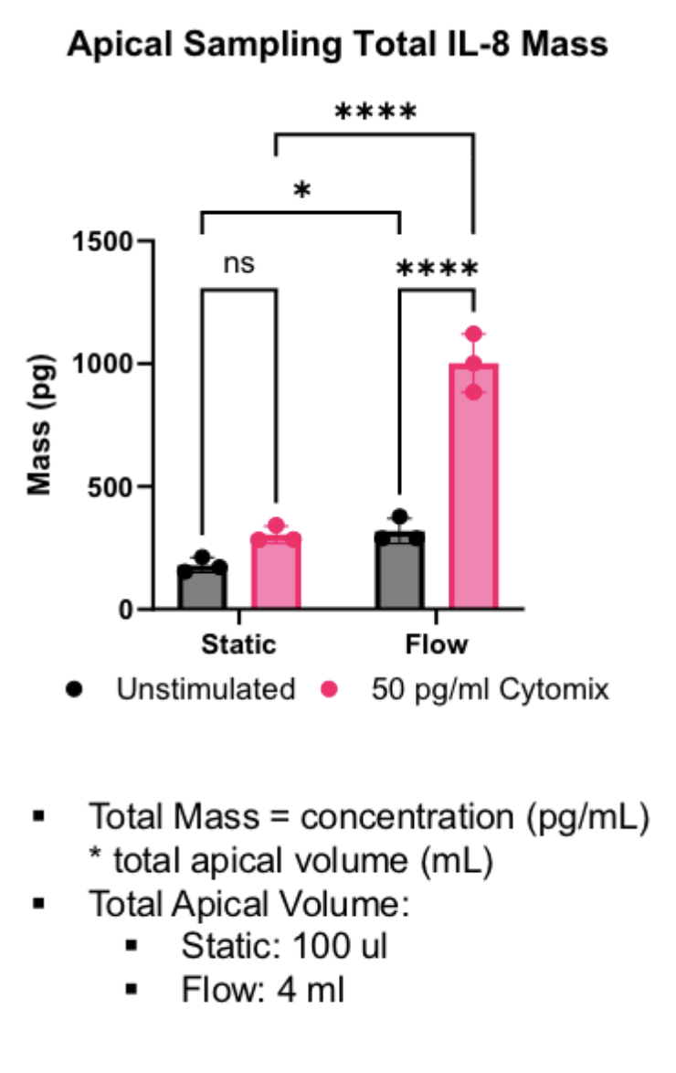
Another interesting calculation is the total mass of IL-8 secreted into the apical side under flow vs. static conditions. It reveals that flow is stimulatory for the secretome response, even though the final concentrations are lower than static conditions.