Flow/No Flow Conditions for Affinity Based Capture of EVs
In previous experiments where the affinity based capture of EVs in ALine devices was tested, issues were encountered. It was noticed that when capturing EVs on the ALine, the non-biotinylated slit membranes had captured a higher quantity of EVs compared to biotinylated membranes. This conclusion led us to question if the flow through the ALine device affected the interaction between the streptavidin conjugated antibody/EV solution and biotinylated membrane surface. To test if flow was an issue for the streptavidin-biotin interaction, we decided to do experiments with flow and no flow conditions for capture of EVs on ALine devices.
Methods
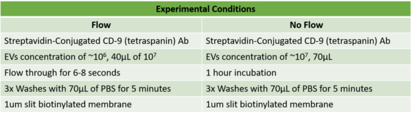
The Hansa lyophilized exosomes from HCT116 cell line were used and prelabeled them with CFSE and streptavidin conjugated tetraspanin CD9. For the ALine devices, only 1um slit membranes were tested. Three replicates of 1um slit biotinylated membranes were used. Table 1 summarizes the experimental conditions ran for flow and no flow. A more detailed protocol on how to coat the membranes and label the EVs, can be found in the following link.
Results
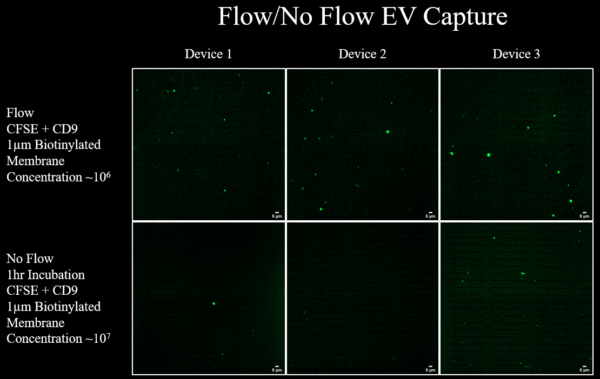
The capture of EVs with the streptavidin conjugated tetraspanin CD9 on the 1um slit membrane Figure 1 looks similar to the results previously discussed, where the 1um slit biotinylated membrane captured much less particles than the amount of particles injected. Figure 1 also shows that flow conditions gives different results, with flow the membrane has more particles captured but it is still a smaller amount than the number of particles injected. The no flow/incubation condition showed almost no particles captured on the membrane.
These results suggested that there might be something affecting the binding of streptavidin to biotin. To test if the avidin-biotin interaction is a problem, 400nm fluorescent streptavidin beads were captured on the 1um slit biotinylated membrane, these results are shown in Figure 2. Since the results from incubating the EVs and not flowing through the membrane, were unsuccessful, the capture of streptavidin fluorescent beads was done via incubation and no flow. As observed in Figure 2, it is clear that beads were successfully captured via incubation. Washing three times after the incubation didn’t affect the amount of beads captured. This suggests that the EV labeling with the streptavidin-conjugated antibody might be the problem.
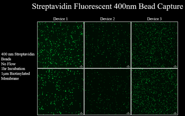
In conclusion, the capture of streptavidin fluorescent beads via incubation suggested that flow is not the problem in capturing EVs. The streptavidin conjugated antibody, in this case the tetraspanin CD9, might not be labeling all EVs since not all tetraspanins are found on the surface of EVs. A more robust antibody would be beneficial for correct labeling of EVs.
Thanks for following through on this Marlyn. Until further notice, I think this spells the end of trying to capture sEVs on slit pore membranes. Works for virus and beads, but we have not been able to find robust conditions for sEVs.