Viability and growth of HUVECs on nanonitride membranes
So I got few nanonitride chips with a pncSi cap on it from Josh (#1019, 320 um sub, 50 nm SiN, 40/20). I annealed them before using it for cell culture. Instead of using the existing CytoVu designs, I made some household cell culturing platforms as shown in the figure below. filled the whole petri dish with cell media (DMEM + 10% FBS + 1% penstrep) till the level of membrane. I am using the frozen HUVEC cells in McGrath lab, currently in P3-P5, for culturing purposes. After multiple murder attempts due to causes like dehydration, depriving cells of gravity, and bacterial contamination, I was finally able to get some stable cell growth on the nanonitride membranes.


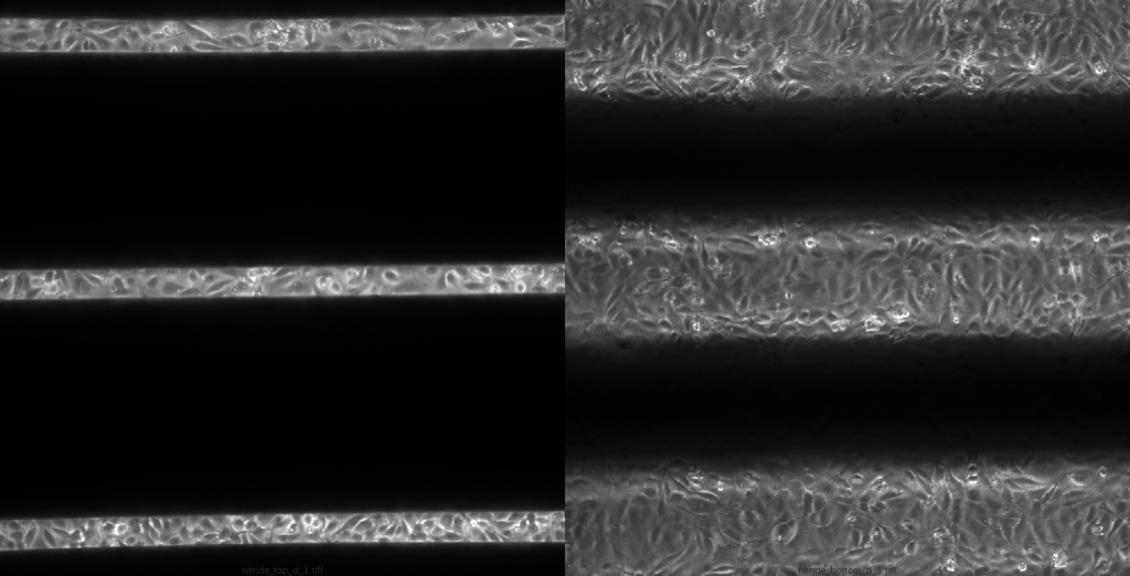
This image shows the presence of cells above the membrane and below the membrane, i.e. on the glass surface.
In order to check whether there are cells on rest of the regions of chips, I stained them with TRITC dye and imaged the chip in inverted position. The staining worked and indeed there are many cells on the windowless regions of membrane as well.
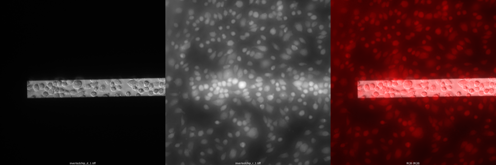
The cells don’t look much spread as they were looking in the previous images. May be the flipping over of the chip caused them to round up. Or may be the staining, washing, and other processes were rough enough to alter their native morphology. Either ways, the aim of the study to show that cells can live and grow on the membrane seems to be satisfies, at least in partial. Not show over here are control images of cells on glass and polystyrene surfaces; cells were >80% confluent on both of them.
Next step is to compare this growth on pncSi, as well as on nanonitride chips with 0 sec and 20 sec RIE, and asking whether those conditions can affect the cell growth or not. I am also planning to perform the LiveDead Assay to look into the cytoxocity of the membranes.