Protein Adsorption on silanized NPN Chip
We are working with the Shestopalov group to silanize the nanoporous silicon nitride (NPN) membrane chip. In an ideal scenario, the silane would then be conjugated to PEG to ward off protein adsorption.
In the presented work, we study protein adsorption on both the untreated and the silanized NPN chips using epifluorescence as the imaging modality. Untreated (n = 3) and silanized NPN chips (n = 3) were incubated (membrane side up) in either 1 mg/mL of FITC-BSA or FITC-IgG for 1 hr, then rinsed/washed in DiH2O and let dry. The NPN chips were then flipped upside down onto a U-shaped gasket for imaging on an inverted microscope (we specifically image the membrane side to match the XPS data). In this study, glass (n = 2) was chosen as the positive control for protein adsorption.
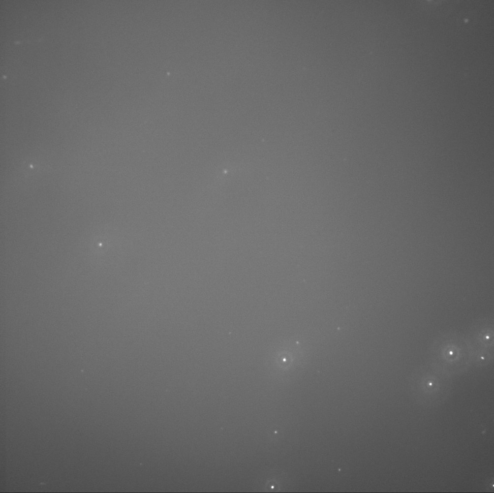
representative image: FITC-BSA on glass:
To keep the imaging locations somewhat consistent, the regions between each pair of adjacent membranes were chosen. Since there are 5 membranes, 4 “between-membrane” regions were imaged for each chip. The schematics below illustrates a representative sampling:

(representative image: silanized-NPN in FITC-BSA)
5 random regions were sampled in the region in between the membranes and 3 random regions were sampled at the left and the right membrane to obtain representative fluorescence values (while bypassing the fluorescence aggregates, which can spike the sampled fluorescence values).
Representative data obtained from each chip:

The broken membranes actually serve as a reference for “baseline/background fluorescence” in this case, which was then subtracted from all data as background correction. The mean value of the baseline fluorescence is quite consistent for all samples, ~80 (arbitrary unit). The fluctuation of the fluorescence is quite high at the membranes, whereas the regions between membranes have similar range of fluorescence. This is true for all the 24 samplings examined (4 regions x 6 chips). Consequently, I only use the fluorescence obtained at the non-membrane regions for the adsorption study.

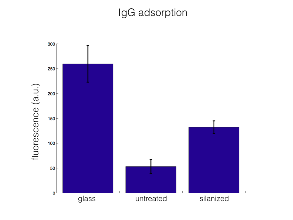
The adsorption of FITC-BSA and FITC-IgG were both higher in the silanized NPN than in the untreated NPN (statistically significant at the alpha level of 0.05/3 using t-test, assuming equal variances).
SIDE NOTE 1: It is quite hard to immerse the silanized NPN into the FITC-BSA and FITC-IgG, indicating that there is a difference in hydrophobocity.
SIDE NOTE 2: I prepared the FITC-BSA and FITC-IgG in house. I still need to perform FRAP studies to assess whether or not the FITC is effectively conjugated onto the proteins.For details please refer to this previous post by Tucker.