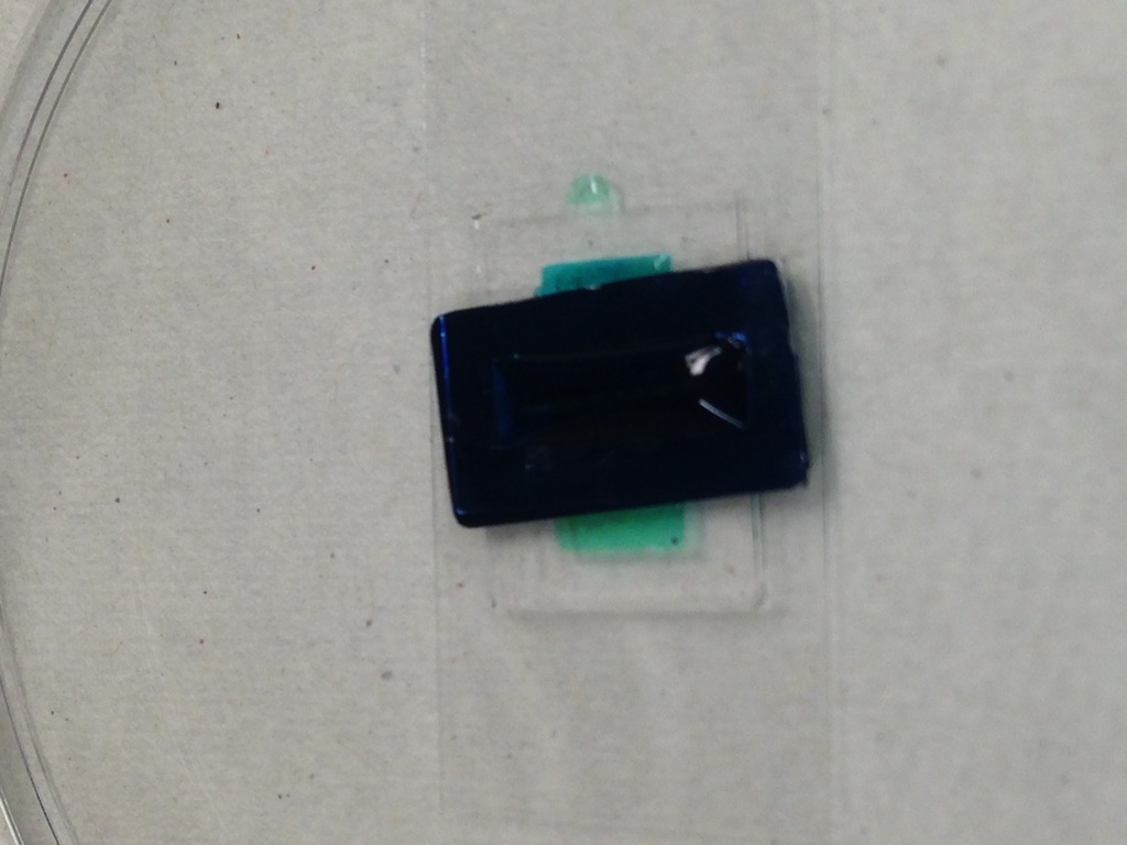
BBB Device V2.8 @ Vaccinex First Seeding Attempts
I made a lot of 6 devices for Vaccinex. We attempted to seed human endothelial cells on the membranes, flowing at 20 uL/min, first coating the surface of the membrane with an adhesion molecule at 20 uL/minute. At the same time, the same cell line was grown in a 24 well TEER z-scope tool to measure TEER (which hasn’t shown any appreciable TEER growth since 5/8/13).
Device 1 (5/7/13)

- Seeded with 20uL/min adhesion factor (basal chamber), then apically with media, then basally with 2e6 cells/mL media. Resulted in sparse seeding.


- 3 ports were clamped with blue ties
- The apical outport capillary leaked, was removed.
- Few cells were seen in the channel, most of the cells formed a monolayer on the ITO glass electrode after 24 hrs of incubation.

- Small air bubbles formed on 5/8, cells still alive. The air bubble was sucked out with a very small syringe needle and replenished with media.
- The tubing was deformed from the clamping, was cut off.
- Cells still around on 5/9, but not really different from 5/8, large air bubbles formed.

- Retired on 5/10, apical channel completely dry


Device 2 (5/7/13)
- Seeded with 20uL/min adhesion factor (basal chamber), then apically with media, then basally with 2e6 cells/mL media. The device was inverted while being seeded, so the flat membrane face was on the bottom side of the basal chamber.
- Basal inport leaked out of the capillary insertion, so cells were injected
- All the cells died within 24 hrs, reseeded with 2e6 cells/mL media.

- The membrane broke after reseeding, not sure when in the process it occured.

- Cells continued to exist in the device, but died on 5/10.
Device 3 (5/10/13)
- Seeded with 20uL/min adhesion factor (apical chamber), then basally with media, then apically with 4e6 cells/mL media.
- Small leak around the apical outport, sometimes self-sealing at the pumping pressures.
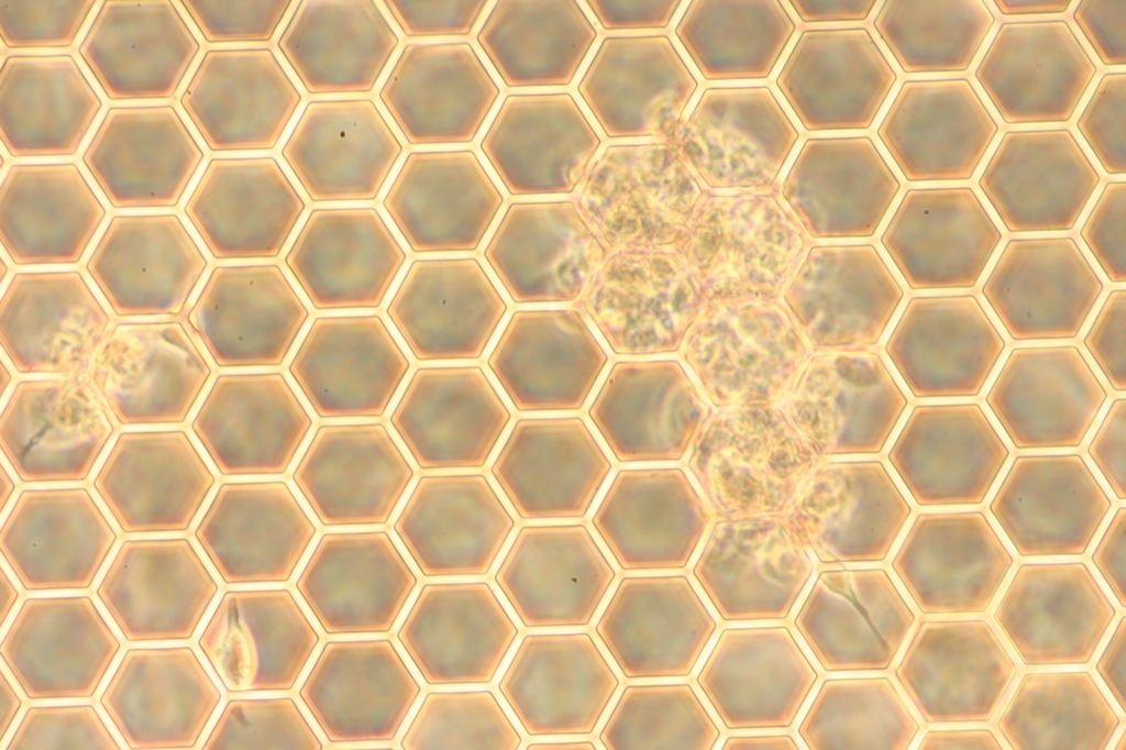
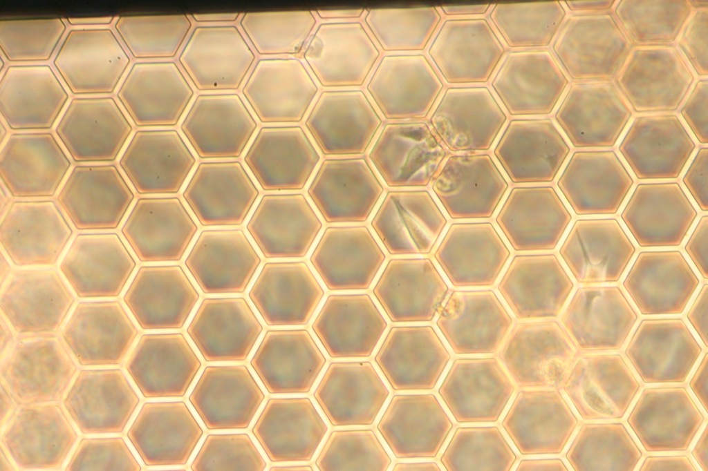
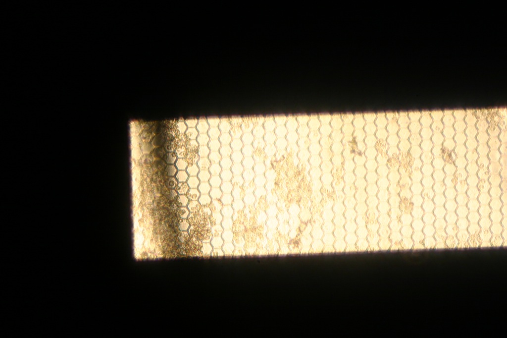
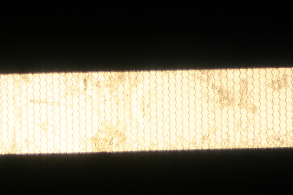
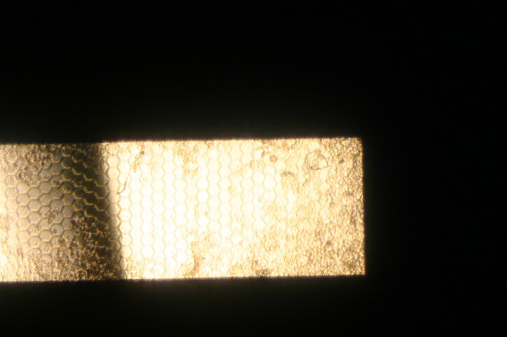
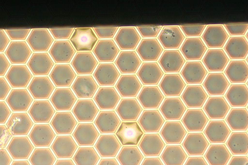
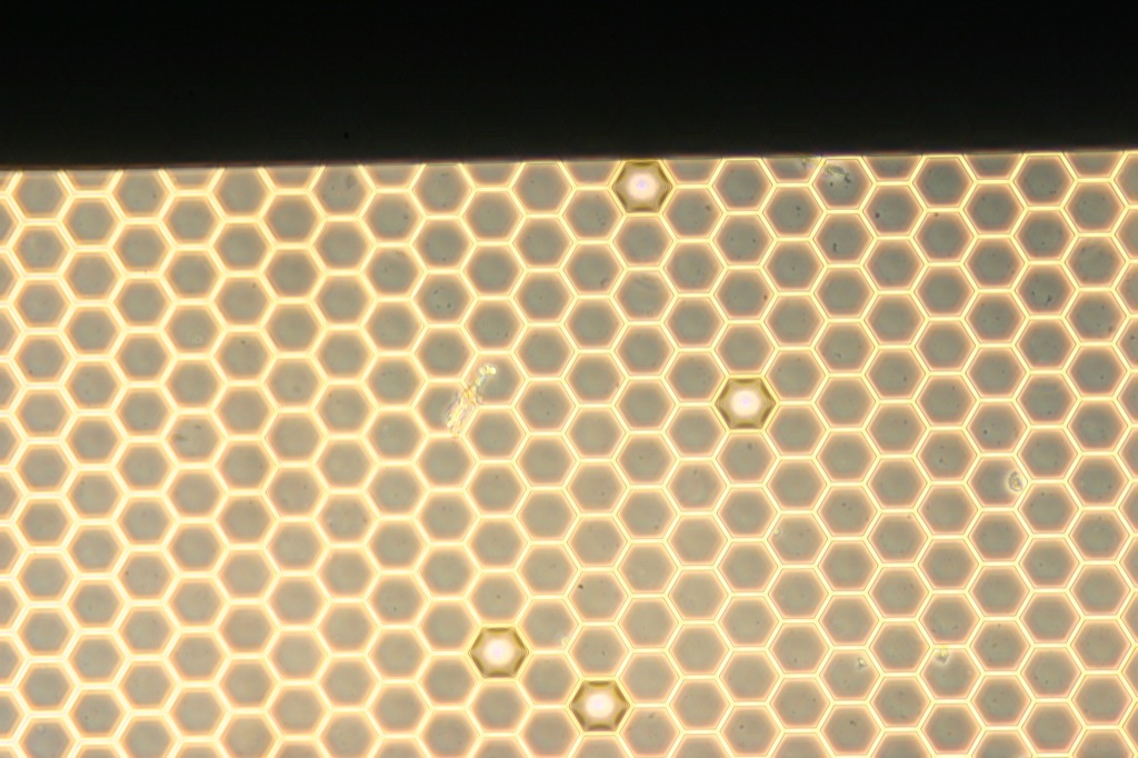
- Large number of cells observed in well on the membrane, clumping especially at the ends of the wells.


- Cells alive on 5/11 with cells extending out of the clumps, as well as isolated cells starting to spread out
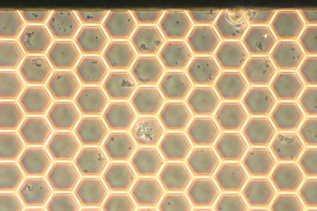
- Cells mostly dead on 5/13. Possible contamination observed on 5/13/13; a fraction of fluid in the channel was collected and will be incubated to check for contamination.
- No air bubbles at all in this device.
Empirical observations
- The total volume for the apical channel (flat side, straight) was ~40-50 uL/device, and the basal channel volume was ~60-80 uL/device. This quantity was established by observing the dispensed volume before entering the glass capillary, and then as the media came out of the corresponding outport capillary.
- Seeding cells on the flat side of the membrane (bottom channel) was troublesome. At the pump rate of 20 uL/min, the cells were settling out of solution before it reached the active chamber. This led to growth on the ITO coverglass electrode face; no cells were sticking to the membrane. Inversion of the channel did not help much either. By cutting away most of the tygon tubing, we were able to minimize the dead volume of the system and get the cells to the membrane better.
- 4 million cells/mL is the cell density that we used to get a large number of cells on the membrane; 2e6 cells/mL was not sufficient.
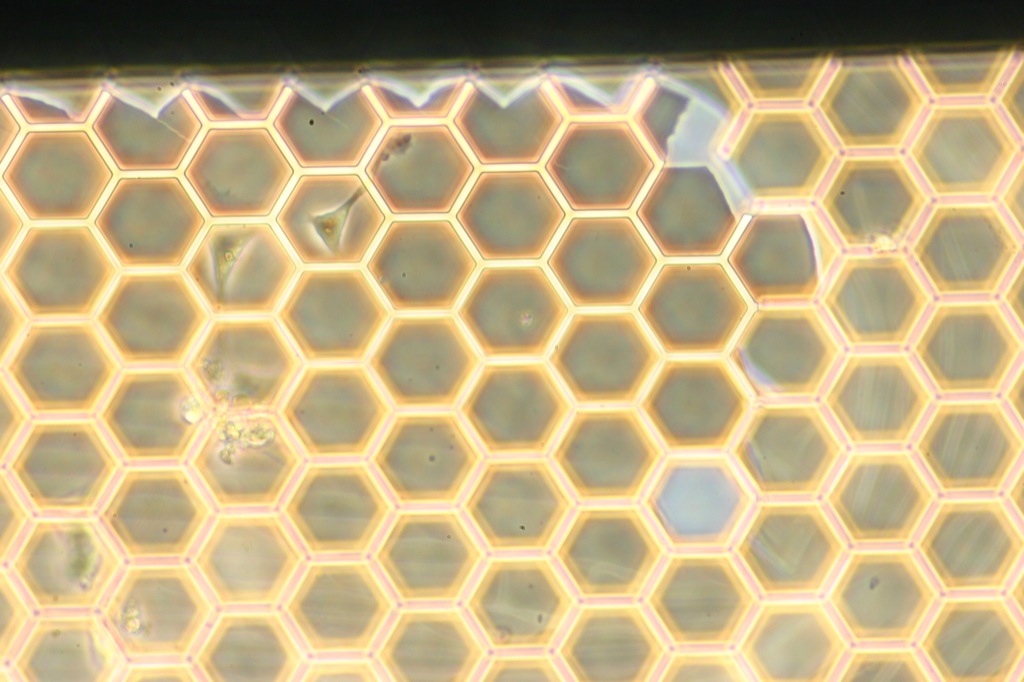
- Removing air is very important to preserving the membrane, but once the membrane is immersed, large flow rates are possible (we were able to move 100 uL of media through a dry apical channel (basal chamber immeresed), in 5 seconds by hand, without rupturing any of the honey comb. Little air bubbles can get trapped in the honey comb.


- Pumping through the channels multiple times with multiple insertions did not fracture the membrane in 2/3 devices, reasonably robust for careful handling. Blunt tip needles were easier to work with rather than the sharp tip needles.
- No leaks between the layers in any of the devices tested; the ozone bonding process has been sufficient for that purpose.
No TEER measurements were obtained from this lot of devices. The EVOM2 either showed a dead short or an open circuit.
- 2 levels of conductive tape did not get the TEER signal to pass after autoclave. However, there was sufficient signal getting to the first level of tape from the ITO, verified with a multimeter. Making the conductive bus out of one piece of tape will rectify this problem.
Next Work
- Determine which adhesion molecules are the best for attaching these cells. Poly L Lysine, Fibronectin are likely candidates. I’ve created 6 open air devices with the single slot dialysis chips (post annealed) to test the cell seeding. The basal chamber is 600 microns high (two 300 micron gaskets), to allow fluid to wick across the silicon into the membrane well

- Determine why the cells die after a few days in the device. Possible reasons include an accumulation of waste products from the cells in high concentration and a lack of attachment. A small amount of perfusion would help remove the waste (less than 1 uL/min) and would not generate a physiologically relevant mechanotransduction stimulus.
- Better capillary insertion/ macro-microfluidic interconnect. All 3 devices had some amount of leakage around the PDMS ports. Jascha’s post could be very relevant here. Alternatively, we may be able to stick the tubing directly in the PDMS feet.
- Get a TEER reading.
- Run a growth run with shear stress.
- More Pictures!