Comparative Single-Well CytoVu Images
After experimenting with some different coatings and membrane types in single-well CytoVu devices, here are some representative images of the results:
SiN (Nitride) membrane with a Geltrex non-gel coating @ 44hr:
10x:

20x:

40x:

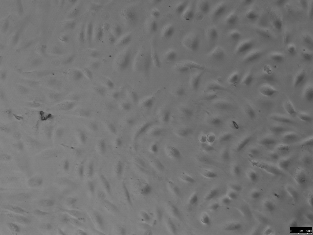
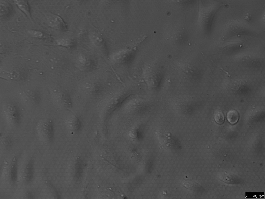
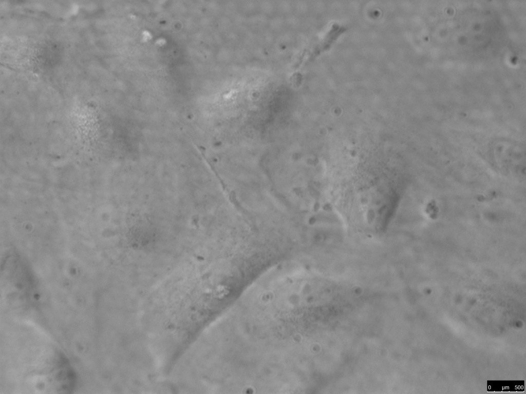
SiO2 (Oxide) membrane with a Geltrex non-gel coating @ 42hr:
10x:
20x:
40x:
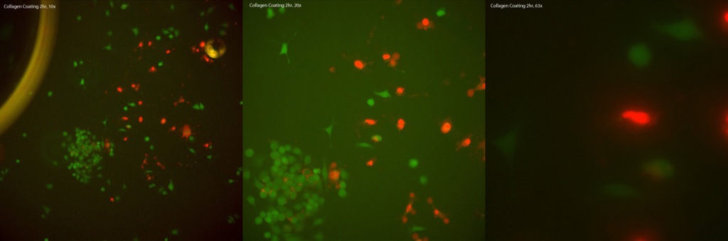
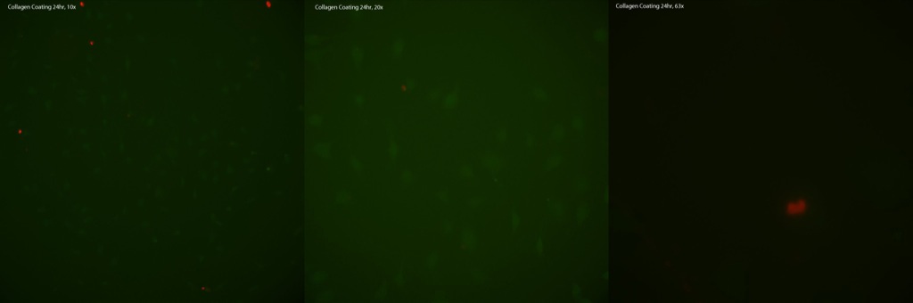
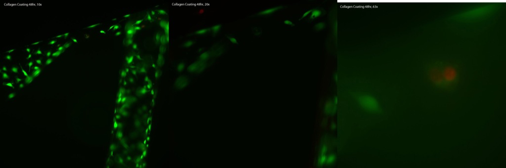
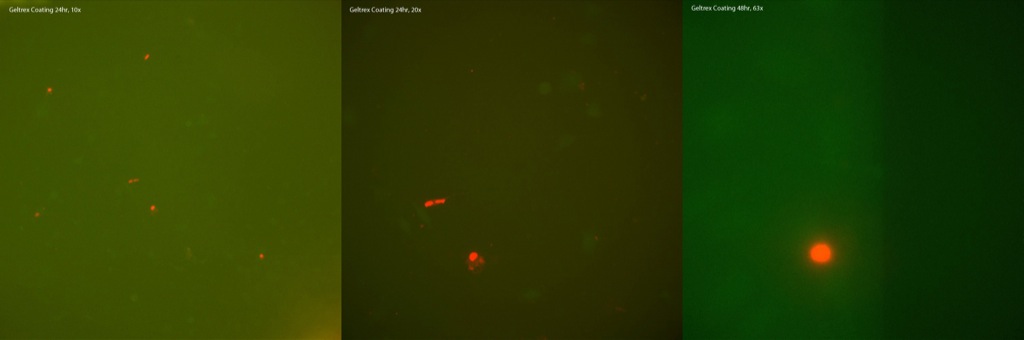
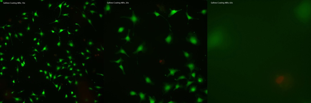
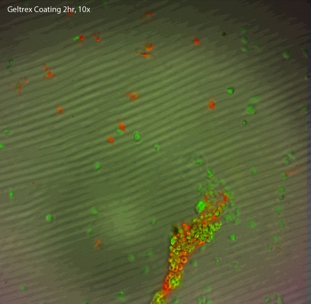
Also, some Live/Dead Assay images were taken on Oxide membranes. These were either coated in collagen or Geltrex (both as non-gel coatings). Representative pictures for each from various timepoints are given.
On both the collagen (first set of images) and the Geltrex (second set), it appears that fewer dead cells were present as the time progressed. This could have been that when seeding, some that were unable to properly interact with the membrane & coating could not survive, but were eventually washed off. Also, since most apoptotic debris would be relatively small, it would likely slip through the 3µm pores and thus would not leave a heavy residue on top of the membrane.
Collagen:

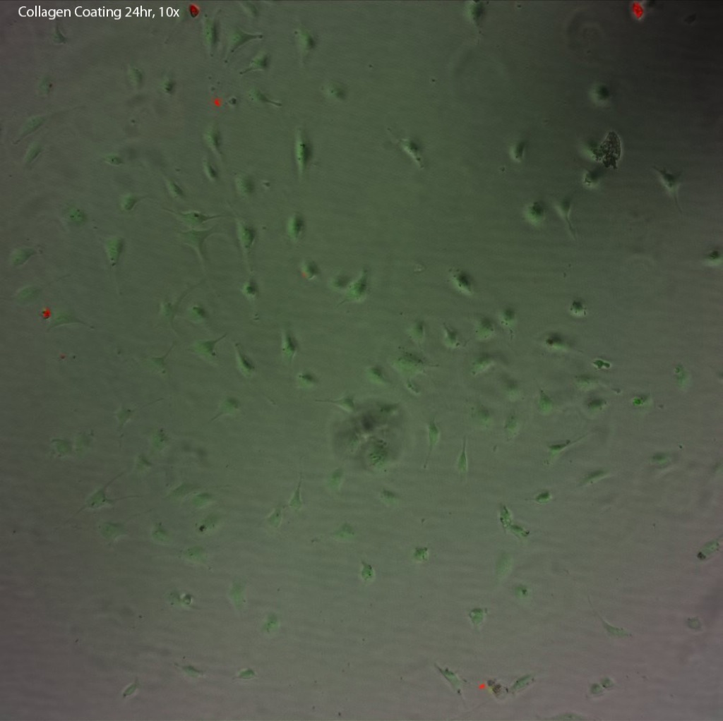
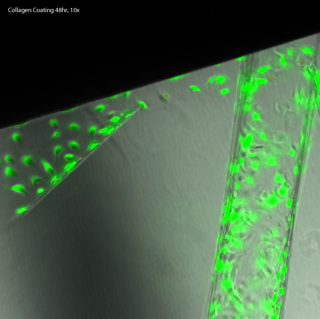
Geltrex:

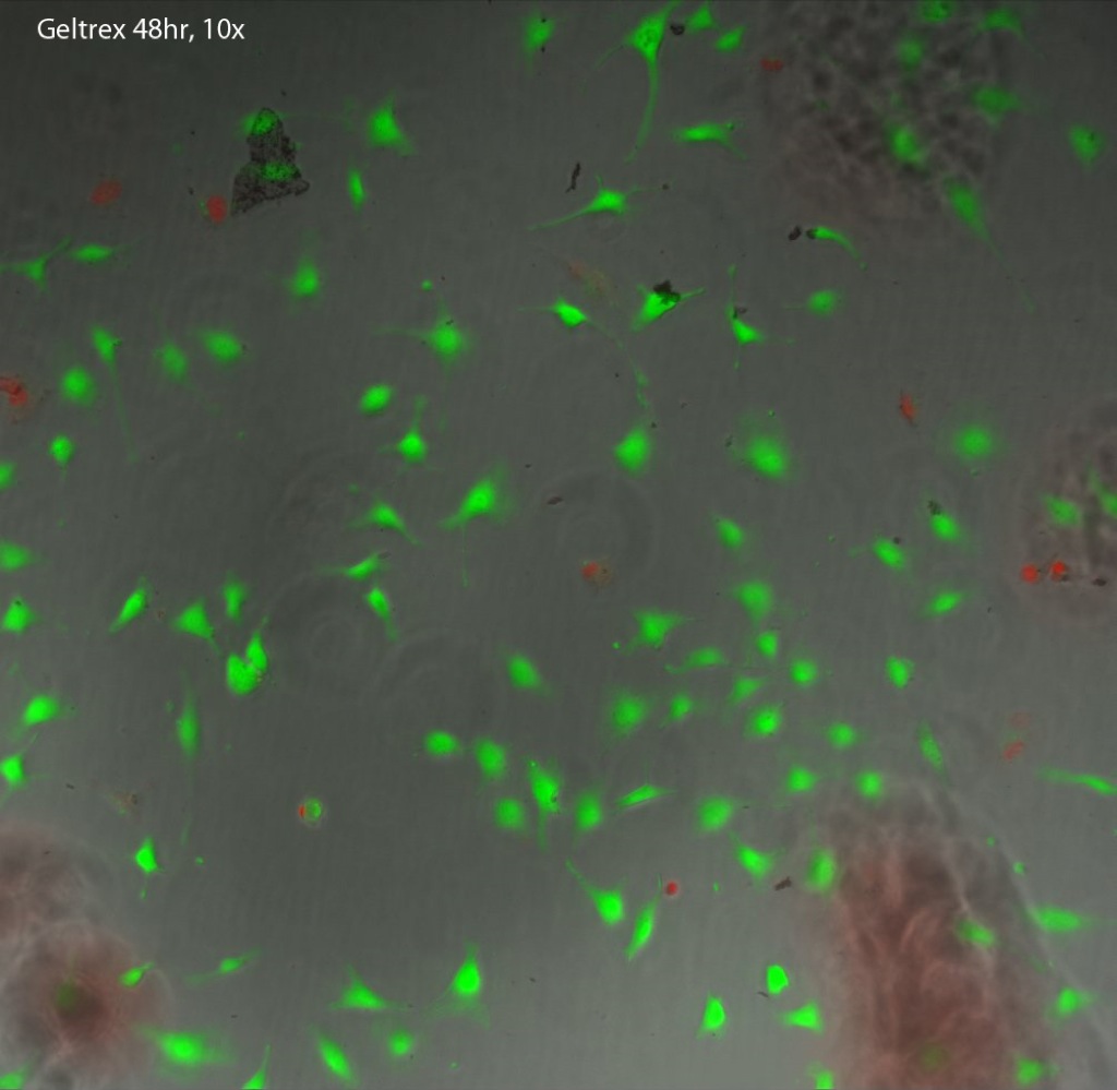
Based on these assays, it would seem that the SiO2 membranes do support cell viability when favorable coatings are placed down on them. Also, due to the optical superiority over SiN membranes, I believe they work better for cell culture purposes (though appear to be somewhat more fragile).
Also, as requested during NRG today, here are the fluorescent-only images from the trial: