HUVEC Cadherin Immunofluorescence (1)
One of the markers of endothelial monolayer maturation/barrier function is the expression of adherens junction and tight junction proteins. The adherens junction is composed of transmembrane proteins called cadherins (among other proteins) that link cadherins of neighboring cells to the actin cytoskeleton. They are found in most (all?) vertebrates and are necessary for cell-cell adhesion in tissues/organs. For tight junction studies, they make a good control protein to stain. Tight junctions are composed largely of the protein ZO-1. Tight junctions are the regulators of paracellular permeability. The more continuous or well-developed the tight junctions, the more impermeable an endothelial/epithelial monolayer is to paracellular transport. Tight junctions have other functions as well.
We have antibodies for ZO-1 and “all” cadherins (pan-cadherin) and different colors of secondary antibodies from Abcam. The protocols are here, here and here. Below are my results from an inital staining study.
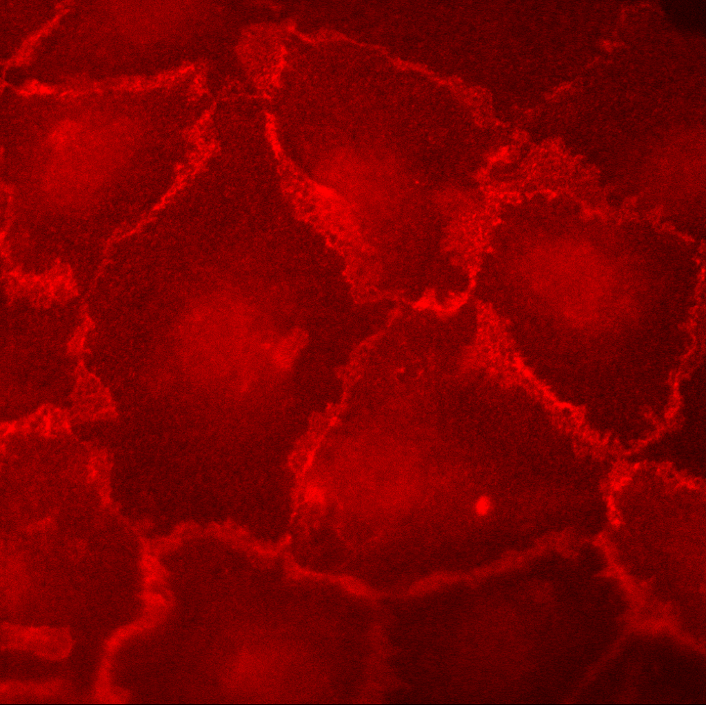
These are HUVEC (P5) on glass cover slips or pnc-Si 7 days post-seeding @ 50,000 cells/cm2. Images were acquired on the Zeiss @ 100X. The pan-cadherin was labeled with a TRITC secondary.
HUVEC on Acid-Washed Glass:
HUVEC on Acid-washed and then PLL-Coated Glass:
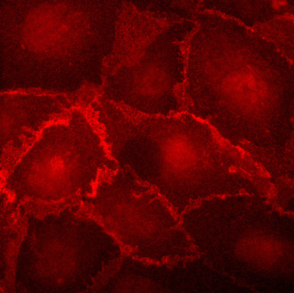
I was interested to see if PLL coating would help cell adhesion/growth, compared to acid-washed glass. It didn’t appear to, which is nice (don’t have to spend the tiime PLL-coating glass cover slips).
HUVEC on pnc-Si:
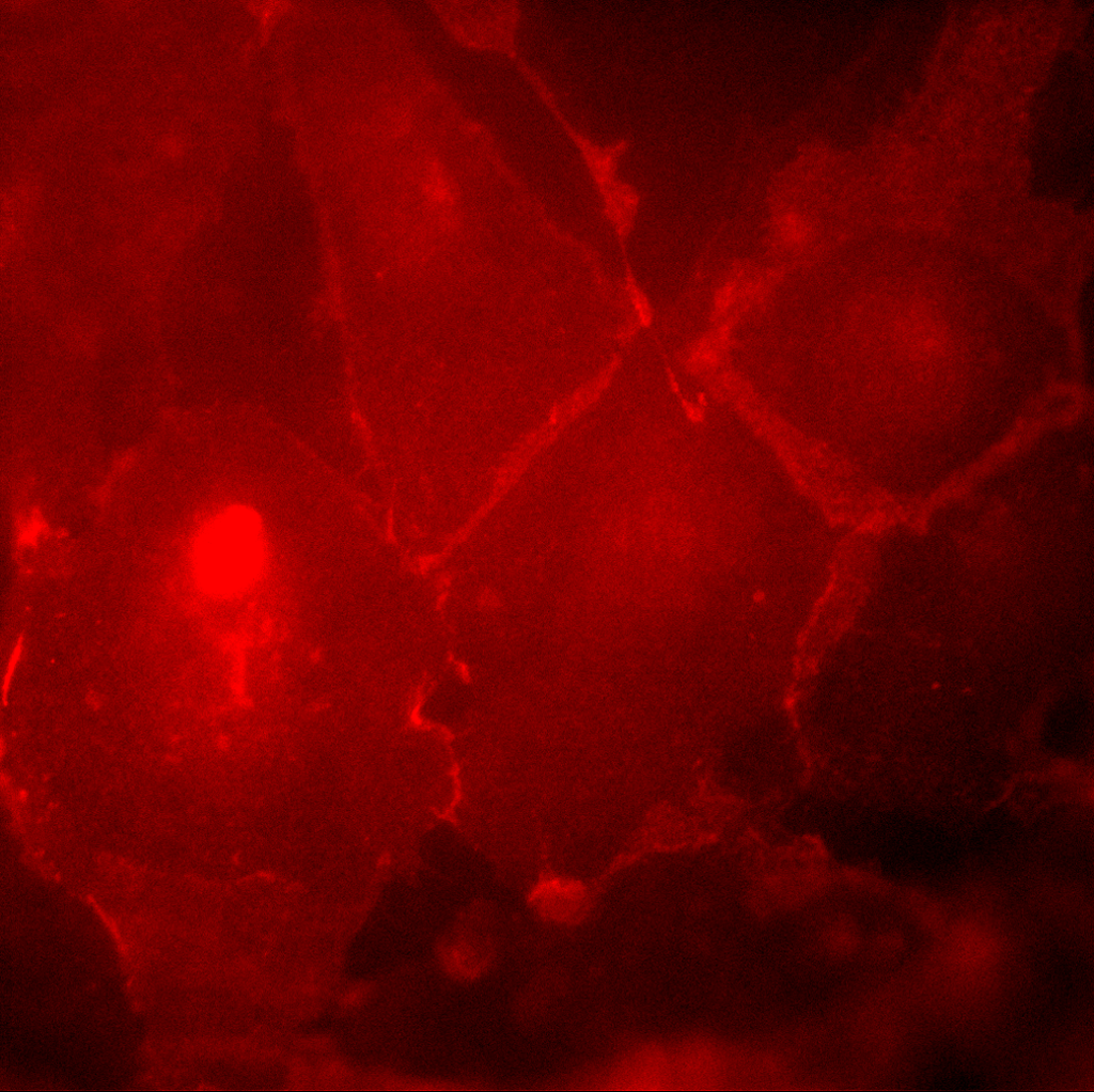
In all samples, there is a fairly high background fluorescence, but cell-cell borders are more brightly stained than anywhere else. This indicates that HUVEC are expressing cadherins at these intercellular junctions (as they should be). Unfortunately, the background fluorescence seemed high, especially on pnc-Si. I need to optimize the protocol for pnc-Si – I was trying to so this procedure with the trnaswells hanging in the 24-wells. I think it might work better if I flip the transwells to do the protocol.
I looked on Abcam’s website and stole this picture of HEK cells stained with pan-cadherin and DAPI from a review of these antibodies:
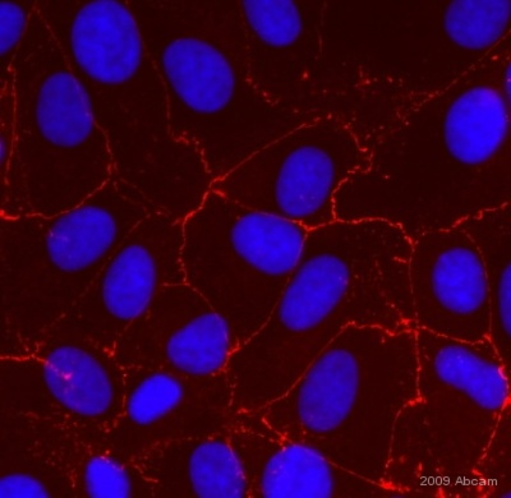 It looks like some background staining might be unavoidable with this antibody.
Have we tried UV-ozone to see if we get better initial adhesion of HUVEC on pnc-Si? I’m concerned that the cells still are not confluent after 7 days on these chips. Do we have evidence that they eventually do become confluent?
i havent tried UV/Ozone but I am using RTP’d samples now since these studies are many days long. I can try next week.