Final BBB TEER results
This post presents the data from all of my TEER experiments of BBB co-cultures, with bEnd3 endothelial cells on the bottom and NG101815 glia on the top of PET and pnc-Si transwells. For all experiments, I used 50000 cells/cm2 as the seeding density. I always had 3 samples – bEnd3 alone, NG10815 alone and the bEnd3/NG10815 BBB co-culture. Passages were between 8 and 17 for bEnd3 and between 8 and 24 for NG101815. Wafer numbers ranged from SC500-613.
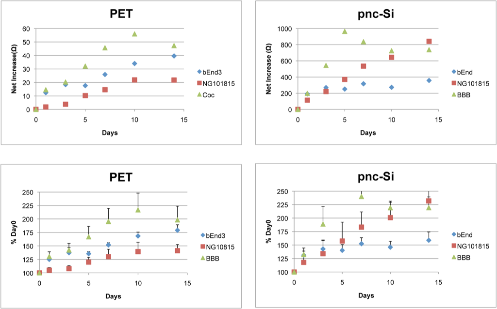
The top row of this graph shows the net TEER increase (in ohms) for each cell type on PET and pnc-Si. For each culture condition (monocultures and co-culture), the net TEER increase on pnc-Si is ~5-20X higher than on PET. The bottom row shows the TEER increase, normalized to the Day 0 TEER (the TEER with no cells on the transwell membrane) – error bars are standard errors. On PET, the bEnd3 TEER increased quickly and then slowly increased over 2 weeks, and the NG101815 TEER slowly increased over 2 weeks. The BBB TEER was super-additive and spiked at day 10. On pnc-Si, the bEnd TEER trend was similar to that on PET. However, NG10815 TEER steadily increased over 2 weeks. This is due to clumping of cells in the pnc-Si wells (remember that these cells are on the well-side of pnc-Si). For the BBB, there is a rapid increase to the spike at day 7 – this appears to be super-additive, as well. However, the NG10815 TEER reaches the same % increase as the BBB at late times, so I think the BBB TEER is dominated by the NG10815 cells after ~ 10 days.
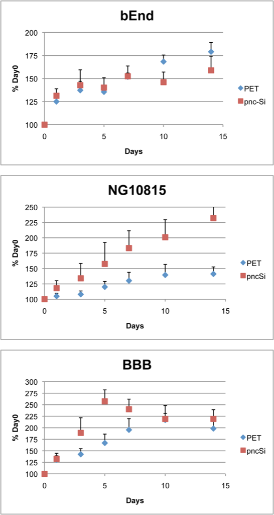
This graphs shows the PET vs. pnc-Si comparison for each culture condition – the same data as above but a little easier to see. Like the ABB results, the bEnd3 TEER on either transwell is about the same. For NG10815 cells, the TEER increases more rapidly, and to a higher maximum, on pnc-Si than on PET. For the BBB, the TEER increases more quickly on pnc-Si and reaches a slightly higher maximum than on PET.
Therefore, there is a super-additive BBB TEER on pnc-Si and PET but it is transient. The TEER for bend3 is similar on both transwells. The TEER for NG10815 cells increases steadily on pnc-Si because of clumping in the wells. This doesn’t happen on PET transwells because the cells aren’t protected in trenches (they get dislodged during handling/feeding). The TEER for the BBB and NG10815 cells increases more rapidly on pnc-Si than on PET. I believe the TEER spike is caused by acidified media. The TEER increases due to glial-endothelial interactions but then the glial cells begin to acidify the media because they are proliferating. This acidification harms the endothelial cell barrier and causes the TEER to decrease after the spike.