Multiplexed digital assay: mouse CCL2(MCP1) CXCL1(KC) panel development
Mouse CCL2/MCP1 and CXCL1/KC pre-equilibrium digital ELISA has been developed with the current dynamic range set to be 4-500pg/ml for CCL2/MCP1 and 8-1000pg/ml for CXCL1/KC. This initial dynamic range was selected by inferring from the ELISA kit reagent working concentration given by the vendor R&D systems (1.9-250pg/ml for CCL2/MCP1 & 8-1000pg/ml for CXCL1/KC) and can be tuned to fit our application by altering the pre-equilibrium incubation time, antibody and enzyme concentration. Two concentrations of detection antibody were tested in the experiment as the increase of detection antibody in CCL2/MCP1 resulting in a marginal effect on the signal strength and an almost two-fold increase in CXCL1/KC, indicating the difference in pre-equilibrium reaction status of the two markers. The current LOD is estimated to reach sub-picogram level for both markers based on previous experience in our sensor background.
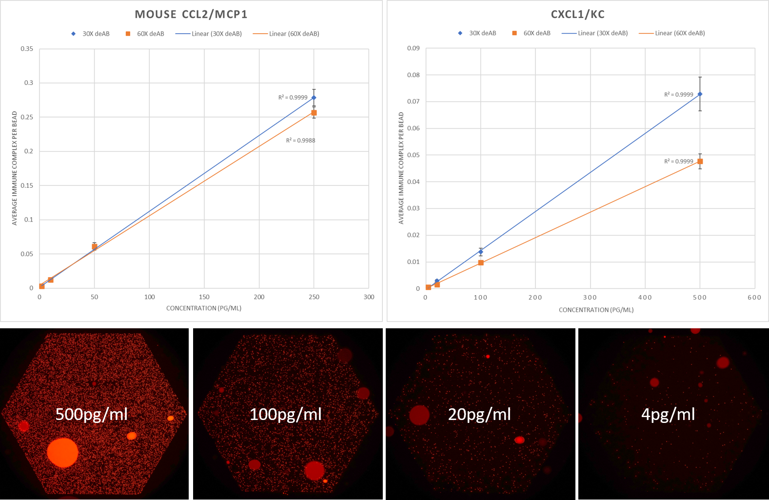
Figure 1. Mouse CCL2/MCP1 and CXCL1/KC calibration curve on 2 detection antibody concentrations and a representative figure of signal output of CCL2/MCP1 in pedELISA sensor array
In order to verify the immunocomplex provided by the vendor is capable of measuring both recombinant protein and native protein. 4 supernatant samples collected from stimulated RAW 264.7(leukemia virus transformed macrophage) cell line was measured. The samples were collected from 96well cell culture plates with a seeding density of 10k cells/200ul and LPS stimulation ranging from 0.8 to 100ng/ml LPS.
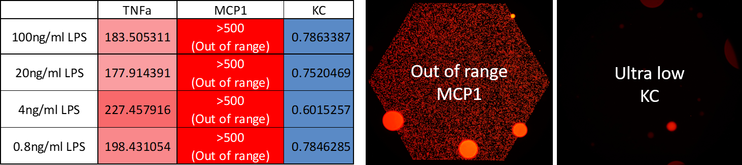
Figure 2. Secretion heatmap of RAW264.7 cells and representative figures of MCP1 and KC sensor output figures from the 4 samples.
The 4 samples secreted around 200pg/ml of mouse TNFa which was similar to our previous experimental range on a per cell level for RAW 264.7 cells. The CCL2/MCP1 secretion level of all 4 samples were all out of range of our highest calibration point which was 500pg/ml. The signal output (average immunocomplex per bead) reached 0.8 which is out of the linear range of Poisson statistics and is thus unreasonable to extrapolate and infer the sample concentration. The CXCL1/KC from all 4 samples were all measured to be under 1pg/ml. Both results match the data from Ref 1. with ultra-high concentrations of CCL2 and not detectable CXCL1 secreted from LPS stimulated RAW264.7 cells. The biological range of endothelial cells and microglia secretion in the uSIM microenvironment can later be assessed in the future and tune our sensor dynamic range accordingly.
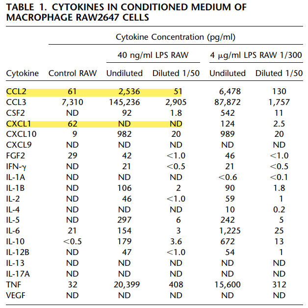
Ref 1. Bein, Di Giuseppe, Mischler, et al.: SFTPB Repression by LPS-Stimulated Macrophage Cytokines, AMERICAN JOURNAL OF RESPIRATORY CELL AND MOLECULAR BIOLOGY VOL 49 2013