Small Particles Task Force: Minimization of Contamination – Current Status (RIT)
The current SPTF team at RIT (Eddingsaas, Hernandez, Routenberg) has been working on blank testing of proposed analysis workflows to extract < 53 µm microplastics from lake water samples, with a goal of ensuring low contamination levels in the workflow prior to analyzing field samples. Field water samples were collected from Lake Ontario throughout 2025 by passing lake water through a 53-µm sieve into a glass sample jar; samples have been archived until < 53 µm analysis methods can be finalized. The theoretical field sample analysis workflow will involve (1) passing the field water sample through a 20-µm mesh (the retentate will be transferred to a filter or kept on single-use 20-µm mesh to analyze for particles from the 20 to 53 µm size fraction), and (2) filtering the permeate (< 20 µm size fraction) through smaller membrane(s) for small particle quantification. Tentatively, we plan to use 8-µm and 1-µm nanomembranes as subsequent size fractions.
Discontinued Blank Processes and Results: Initial 8-µm nanomembrane blanks in 2025 which attempted the entire workflow had large fibrous contaminants that were reduced by switching from a reusable sieve to custom cut 20-µm disposable steel mesh filters (Routenberg). In early 2026, blank attempts using water filtered with 0.8 µm mixed cellulose ester (MCE) filters continued to have higher levels of contamination than desired (Fig. 1, Hernandez). These blanks implemented furnace ashed glassware. Blank tests stepped through different parts of the proposed workflow, progressing towards the goal of transferring water passed through 20-µm disposable steel mesh filters onto smaller size fraction nanomembranes. However, results worsened as more steps and transfer processes were added to the workflow (i.e., R1 to R3 from left to right in Fig. 1).
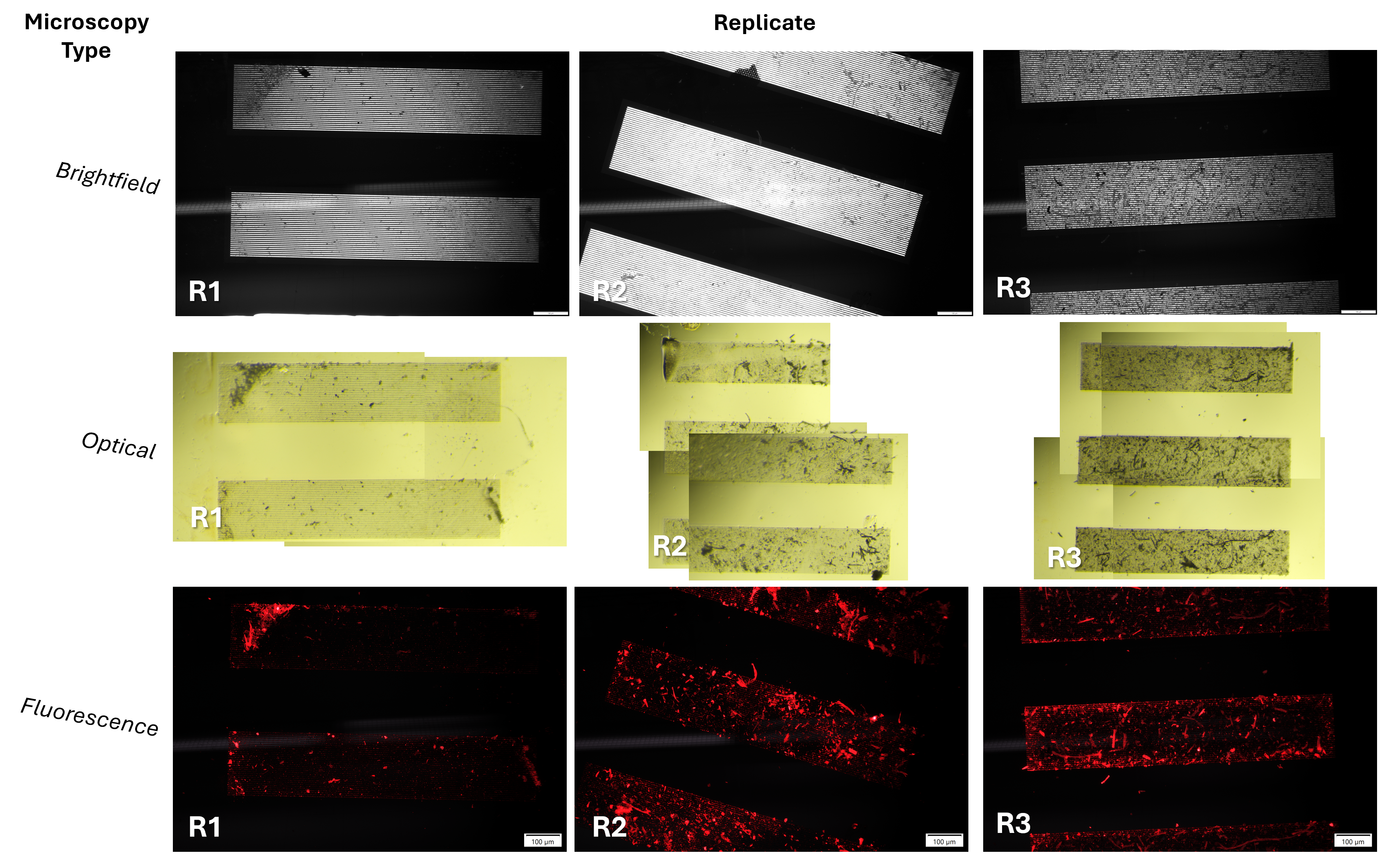
Following these results and discussion with the group, MCE filter use was discontinued (suspected to shed contaminants into 0.8 µm filtered water) and subsequently replaced with syringe filtration. Furnace ashing of glassware was also discontinued as an unnecessary handling step that could worsen contamination, based on feedback from the larger SPTF group. Recent blanks use the cleaning procedures described below.
Current Cleaning Procedures: Techs always wear 100% cotton, pink lab coats and gloves that are rinsed with ethanol as described in the MMC minimization of contamination blog post. Glassware is cleaned with natural fiber sponge and soap, followed by 3x DI rinse, followed by 3x filtered DI rinse. For small particle samples, a final 3x rinse of glassware is done with syringe-filtered water (using sterile 0.45 µm SFCA membrane). The syringe filter is prepared by flushing at least 60 mL of DI through the setup before using it for rinsing or sample processing steps, to avoid introduction of contaminants shed when the syringe filter is used for the first time. The final glassware rinse and further sample processing steps are performed inside a Biological Safety Cabinet (BSC), which is first cleaned by wiping 3x with 70% EtOH.
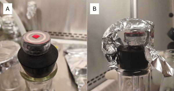
Recent Blank Processes and Results: Recent blanks use 50 mL of syringe-filtered DI water as the blank solution and syringe-filtered DI to rinse the vacuum filtration funnel. We tested filtration of Nile Red (NR) solution and isopropyl alcohol (IPA) through both syringe filtration (Fig. 2, Test 1; note that acrylic syringe filter housing broke after exposure to alcohol) and through the 1 µm nanomembrane (Fig. 2, Test 2). For this series of tests, we also imaged the nanomembranes between blank water application and staining, determining that NR/IPA solutions seemed primarily responsible for introduction of contaminants (Fig.2).

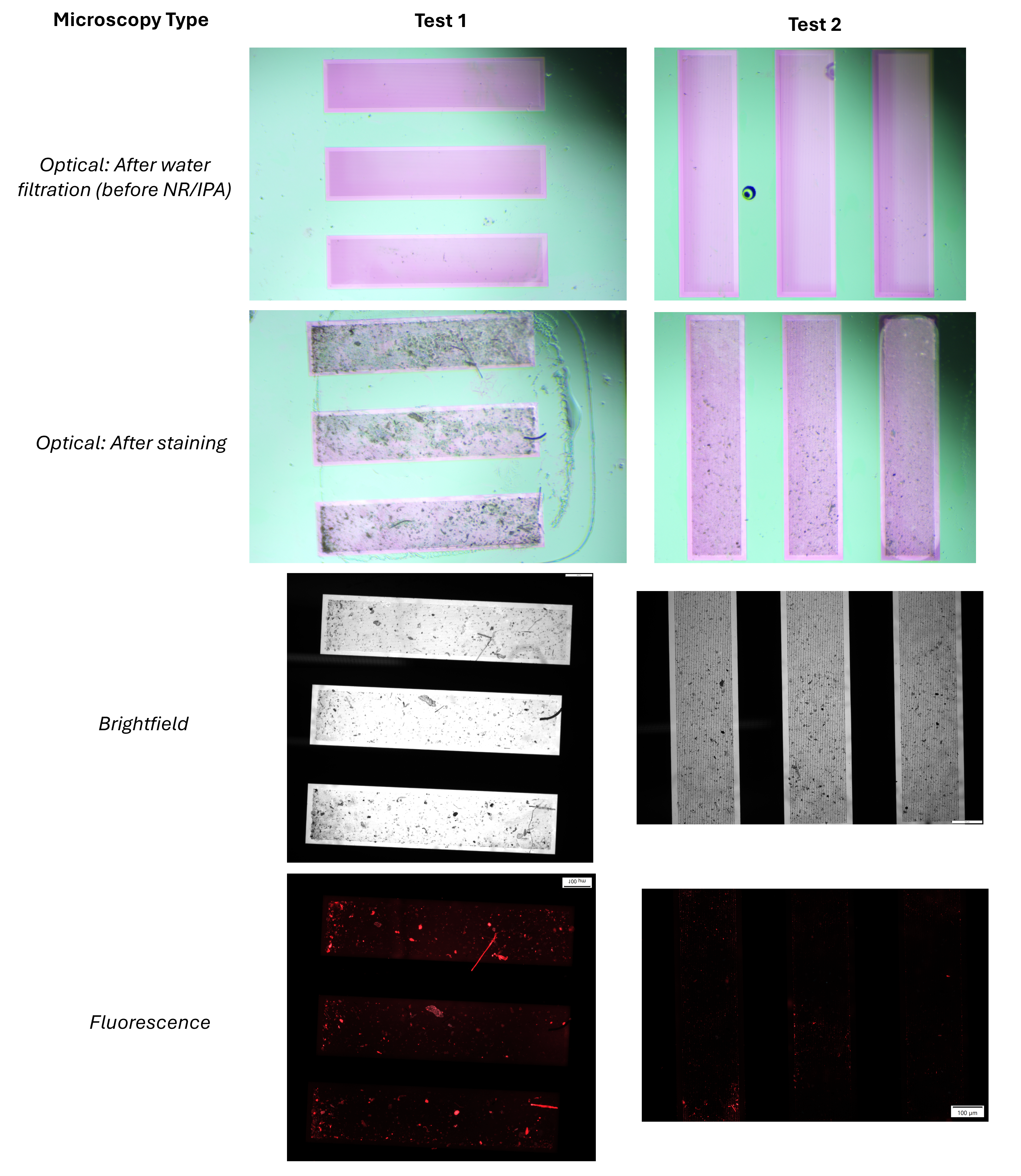
Next Steps: Nanomembrane filtration of NR/IPA seemed to be successful in reducing blank contamination. However, this method is expensive with our current prepared solution (several nanomembranes were used to filter NR/IPA solutions for the results of Fig. 2, due to clogging issues). Our next steps are to prepare a new high concentration Nile Red stock solution and try filtration through a 0.5 µm nanomembrane (via sepcon), then use the stock to create less concentrated solutions for further blank testing; this method is used successfully by others within the larger SPTF group.