HUVEC morphology recovers on pnc-Si
Since HUVEC morphology hasn’t looked good in short-term adhesion assays recently, I analyzed HUVEC on the 2nd day after cell seeding.
Normally, CMFDA staining is done for 30-45 minutes in serum-free media. I wondered if the serum-free environment during staining was affecting the HUVEC. So, I stained one sample with 5uM CMFDA in serum free media and 1 sample in 10uM CMFDA in serum-supplemented media. These are commercial PET transwells.
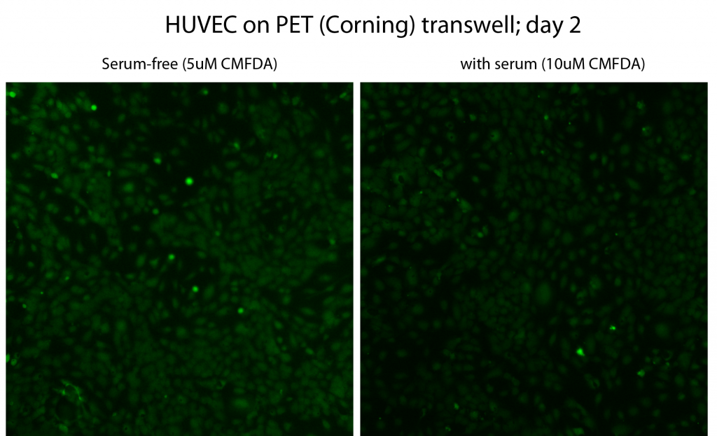
It looks like the staining in serum-free media is brighter, which isn’t surprising. Molecular Probes recommends serum-free media because CMFDA amines and thiols inactivate the CMFDA. I tried to get around this limitation by doubling the [CMFDA]. It does appear that high CMFDA concentrations do work in sera-supplemented media, which is important if we ever work with very sensitive cells. I’ll update the protocol. HUVEC mrophology is normal, similar to that after 5 hours (here).
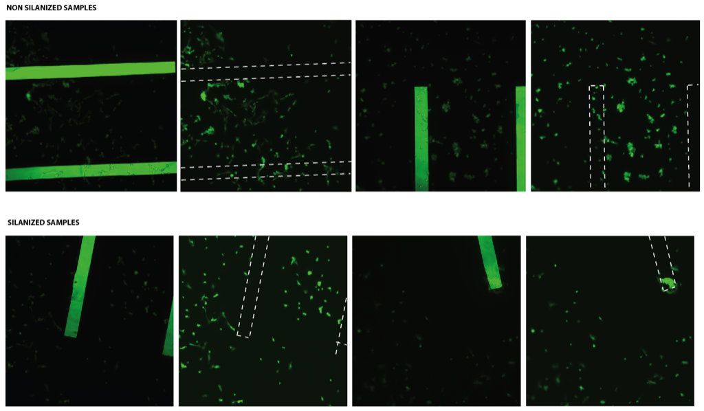
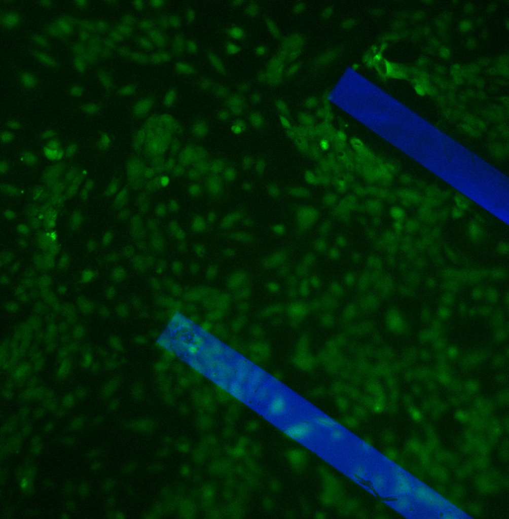
HUVEC morphology has not been normal on pnc-Si after 5 hours lately (here, here also). By contrast, HUVEC morphology looks good after 2 days in vitro (membrane slits in blue):
This agrees with recent data posted by Anant (here, here-kind of).
I’m not sure when the top membrane broke here, but the HUVEC have arranged themselves around this slit. If the membrane broke during staining, the fluid could have just pushed those HUVEC out of the way. If the membrane was broken for the whole culture period, HUVEC may be reacting to the edge of the membrane slit.
The main conclusion from this study is that it takes 1 or 2 days for HUVEC to adjust to pnc-Si and elicit a normal morphology.