Focal Adhesion Formation on PDMS Substrates
Background
Focal adhesions occur in 2D cell cultures as substrates apply large forces on cells. Previous experiments showed that focal adhesions formed on TCP and on non-porous SiO2 membranes but that less formed on 3.0µm-pore SiO2 porous membranes and even less on 0.5µm-pore SiO2 porous membranes. In other words, as area between pores decreased, formation of focal adhesions decreased. Our previous experiments concluded that HUVEC respond to non-porous SiO2 membranes as stiff, 2D substrates, but respond to 0.5µm SiO2 membranes similar to compliant, 3D matrices. In this experiment, HUVEC were seeded on top of two different types of PDMS substrates with varying stiffness to analyze focal adhesion formation on soft substrates.
Methods
- A 24-well plate was prepared with the following substrates:
- TCP (4 wells),
- Sylgard 184 (4 wells) – 400mg/well
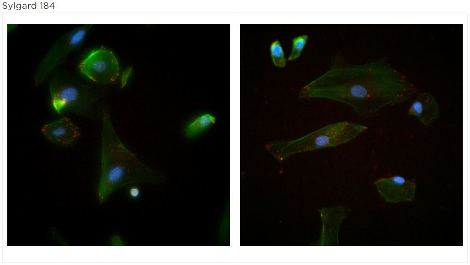
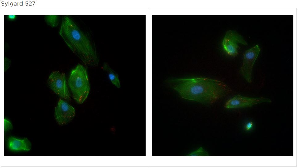
- Sylgard 527 (4 wells) – 400 mg/well
- 300µm-thick square PDMS gaskets were cut using the craft cutter with outer dimensions of 5.65mm x 5.65mm and inner dimensions of 3.75mm x 3.75mm, creating seeding area of 0.16cm2. The gaskets were adhered to the substrates using ozone treatment. The plates were cured overnight and exposed to UV light for 15 min. on each side before seeding cells.
- Each gasket was coated with 1% Geltrex.
- HUVEC were seeded at 5000 cells/cm2 inside each gasket.
- After 24 hrs, the cells were stained with fluorescent-labeled DAPI to stain the cell nuclei, Phalloidin to stain actin filaments, and monoclonal antibody to Vinculin to stain gap junctions.
- Wells were imaged using the DAPI, GFP, and TxRed fluorescent channels.
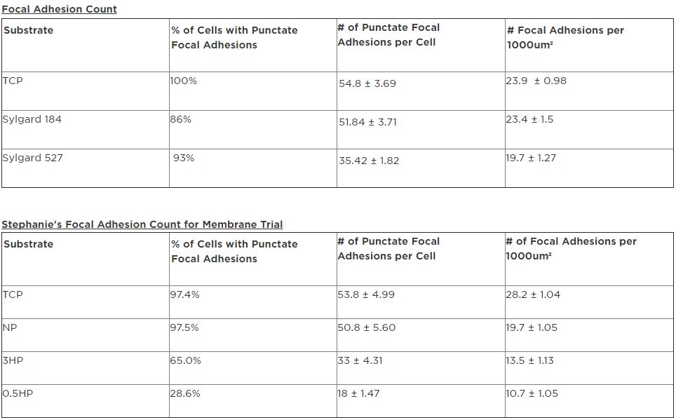
- After obtaining the images and overlaying them, punctate focal adhesions were counted and recorded for comparison.
Data

Conclusion
TCP values between this trial and the membrane trial were compared. The values were consistent, indicating that the analysis performed is valid. Sylgard 184 (E= 1.72MPa) had focal adhesion values comparable to the Non-porous membrane. Sylgard 527 (E= 5kPa) had focal adhesions comparable to the 3µm-pore high porosity membrane but the latter had less % of cells with punctate focal adhesions. These results are consistent with what we hypothesized – that the cells on the softer substrate (Sylgard 527) would have less focal adhesions than on the stiffer substrates (Sylgard 184 and TCP).