Microtechnologies for cells an devices – BMES 2019
Microtechnologies for cells
Phototunable Spatiotemporal Presentation of Viscoelastic and Adhesive Cues in HA Hydrogels
Erica Hui1, Kathryn I. Gimeno2, Thomas H. Barker2, Steven R. Caliari1,2
Department of Chemical Engineering, Department of Biomedical Engineering, University of Virginia
https://www.biorxiv.org/content/10.1101/646778v1.full
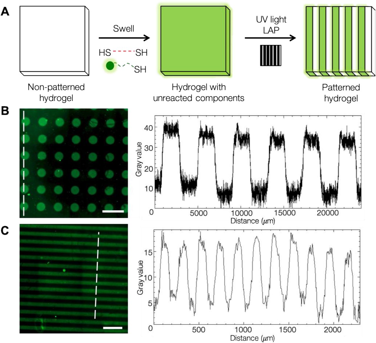
Developed a hyaluronic acid (HA) hydrogel system to study the role that spatially-patterned matrix mechanical (viscoelastic) and adhesive properties play in regulating cell mechanotransduction and myofibroblast activation.
Photopatterned hydrogels were fabricated through swelling an initial hydrogel in a solution of crosslinker, photoinitiator, and thiolated fluorophore
with a photomask to regulate UV light penetration.
Photopatterned regions produced elastic moduli (E) comparable to stiff fibrotic tissue (E = 14.20 ± 2.56 kPa) and viscoelastic regions displayed frequency-dependent and stress relaxation behavior not seen in elastic regions. Cells responded to the local mechanics of the patterned substrate with increased spreading in fibrosis-mimicking regions (stiff regions).
Dissecting Nanotopographical Regulation of Cell Behaviors
Kai Wang, Kun Man and Yong Yang
Department of Biomedical Engineering, University of North Texas, Denton, TX 76207
Nanotopographical and biochemical cues were decoupled as shown in Figure 1a. Nanogratings (650 nm in linewidth, 500 nm in spacing) and nanorectangles (650 nm in length, 500 nm in width) were fabricated on polystyrene (PS), which cells did not deform. Rhodamine-labeled FN (rFN) nanopatterns (biochemical cues) were stamped on the surfaces of these substrates.
Nonflat surfaces promote alignment and less spreading, also have more focal adhesions as shown by fibronectin, reduced proliferation, reduced yap ratio, and laminin accumulation inside the trenches. Authors concluded that both nanotopographical and biochemical cues affect stem cell behaviors,
but the nanotopography has a more profound impact than the biochemical cues.
Question: what is the limit in size that will still have an effect? cell-type dependent but metastatic cells can probably sense smaller features

Microtechnologies for devices
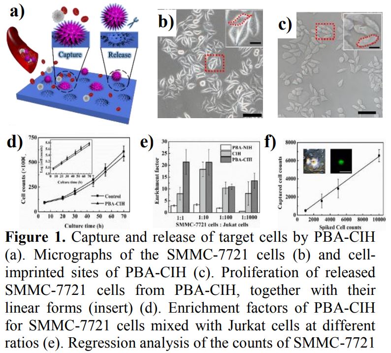
A Cell-Patterned Hydrogel with Hierarchical Structures and Boronate Affinity: Towards the Selective Capture and Undamaged Release of Tumor Cells
Kaiguang Yang†,1, Lukuan Liu†,1, Lihua Zhang*,1 and Yukui Zhang1
Dalian Institute of Chemical Physics, Chinese Academy of Sciences, Dalian, 116023, China
Human hepatocarcinoma SMMC-7721 cells were used as the tumour cell templates. They were first adhered and adequately stretched in the culture dish. Then, the functional monomer, the crosslinker and the affinity phenylboronic acid monomer, 3-AAPBA, were added to form a hydrogel on the cell templates. Finally, the hydrogel was peeled off with the imprinted face up, and the cell templates and unreacted monomers were removed by trypsin and deionized water to fabricate pre-designed biomimetic interfaces with the boronate group exposed on the hydrogel surface.
SMMC-7721 cells in amounts ranging from 10^3 to 10^4 were spiked into 1 mL of whole blood. As shown in Figure 1f, a linear correlation between the counts of spiked and captured cells (R2 = 0.997, n = 3) was observed, the capture efficiency of SMMC-7721 cells was 49.9 ± 14.5% (n = 3)
Device Fabrication Technologies for Protein-Based MEMS Implants
Author: Mark G. Allen, University of Pennsylvania
Recently it has been hypothesized that implants microfabricated from materials such as extracellular matrix (ECM) protein, could offer the possibility of MEMS-based devices that are more easily integrated with the body, are better tolerated by the immune system, and/or which have mechanical properties that are well-matched to potential biological applications. However, the incorporation of these relatively delicate materials into standard MEMS processes can result in significant fabrication challenges since proteins are temperature/chemical/light sensitive.
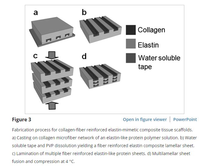
Example 1: Aligned collagen fibers embedded in elastin.
https://onlinelibrary.wiley.com/doi/full/10.1002/adhm.201300112

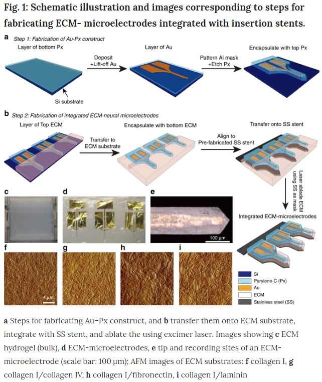
Example 2: Neural electrode encapsulated in ECM (collagen, fibronectin and laminin)
https://www.nature.com/articles/s41378-018-0030-5
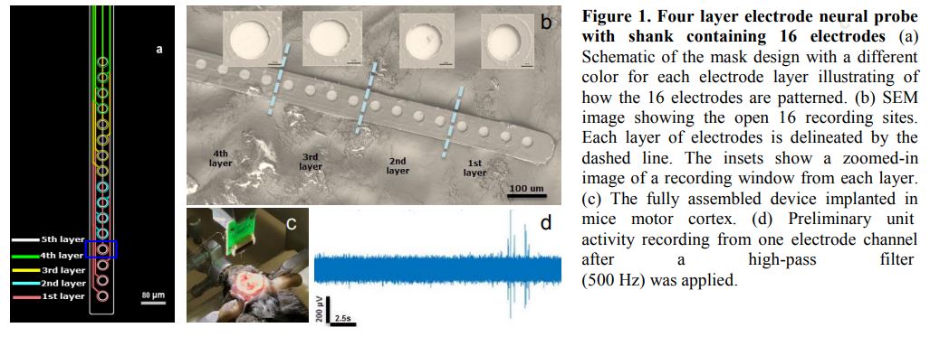
Multilayer Implantable Cortical Microelectrodes to Improve Recording Potential for Spinal Cord Injury Treatment
Xin Liu1, Elena Bibineyshvili2, Fernando Rebolledo1, Sunshine Littlecreek1, David J. Margolis2, Hilton M. Kaplan3, Joachim Kohn3, David I. Shreiber1, Jeffrey D. Zahn1
1Department of Biomedical Engineering, 2Department of Cell Biology and Neuroscience
3New Jersey Center for Biomaterials, Rutgers University, Piscataway, NJ
Presented a methodology for scaling up the number of recording sites without proportionally increasing the size of the probe by patterning recording
electrodes vertically on multiple Parylene C support layers. The electrodes are used for brain/computer interfaces. Fabrication of the multilayer probe involves repeated photolithographic definition of titanium/platinum (Ti/Pt) electrodes and chemical vapor deposition (CVD) of Parylene C. Reactive Ion Etching (RIE) was adopted to define the final probe geometry and open recording sites.