Investigating vacuoles on transwells
One of the themes in my recent posts has been the presence of ‘vacuoles’ over free-standing pnc-Si and PET membranes. We’ve assumed these are vacuoles because endothelial cells form vacuoles, they don’t stain with live cell stains (and so have no cytoplasmic enzymes) and there can be several of these structures in each cell. In the past month, I’ve done a couple of experiments to try to establish if these structures are indeed vacuoles.
First, I used a blue-fluorescent Lysotracker Dye from Invitrogen to see if these structures are lysosomes. This dye is membrane-permeant and tends to accumulate in acidic organelles (like lysosomes) but the exact method of action is unknown. Here, I incubated cells in 75nM Lysotracker (in growth media) for 2 hours, replaced with dye-free media and observed at 20X on the Zeiss. I counterstained with 10uM CMFDA for 1 hour to label live cells green. These are P12 bEnd3 cells grown for 1 days on SC500.
PET:
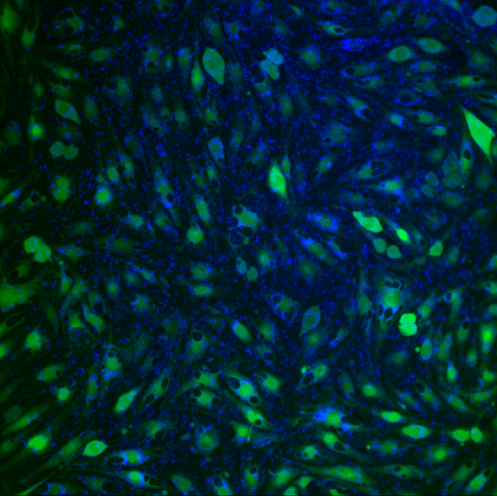
pnc-Si:
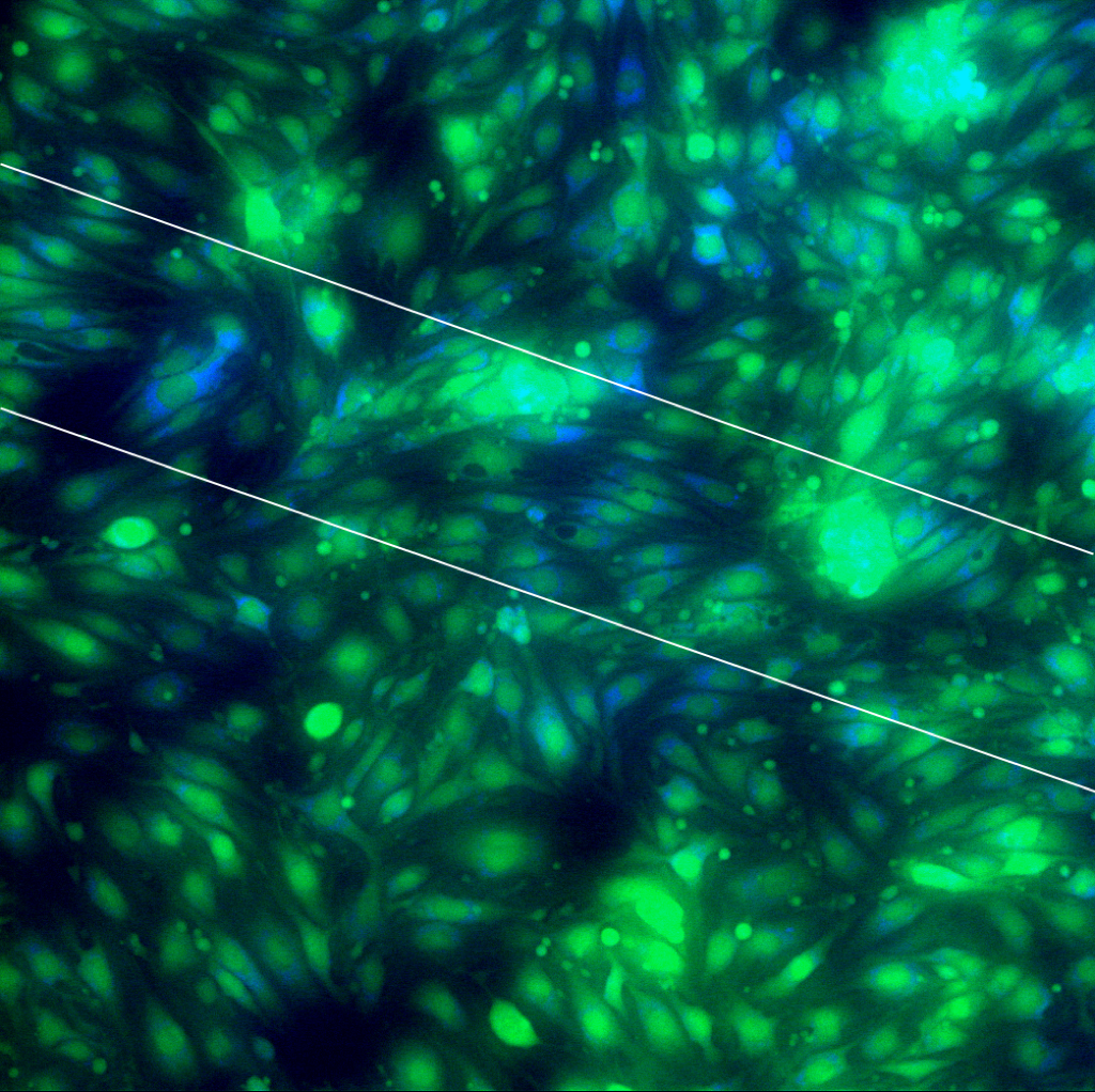 The Lysotracker staining wasn’t very bright on either of these samples (the dye wasn’t stored properly and it might be bad), and the blue channel looks a little washed out. Also, it seems like the dye stained the pores of the PET sample. Apart from background fluorescence, it looks like the dye concentrates in perinuclear regions. Importantly, I don’t think there is an instance with blue fluorescence inside a black (non-green stained) vacuole on either sample. If this dye is still OK, these images suggest that the ‘vacuole’s are not lysosomes. I think if we want definitive proof, we need to buy new Lysotracker (or similar) dye.
Another experiment I tried was inspired by the Nature paper I reviewed for journal club a few months ago. In this paper, they labeled vacuoles in vitro and in vivo by incubation with Dextran-Tetramethylrhodamine (3kDa) at 0.1mg/mL (or 2ug/mL carboxytetramethylrhodamine). The idea (as I understand it) is that membrane-impermeant dyes that aren’t ligands for receptor-mediated endocytosis should only enter cells via pinocytosis and end up in fluid-filled vacuoles. The closest dye we have to tetramethylrhodamine is tetramethylrhodamine-5-iodoacetamide dihydroiodide (TMRIA).
For these images, I seeded bEnd3 cells, allowed them to attach for 2 hours and then let them grow in dye-supplemented media for 1 day. I used 2 dyes – green FITC-Dextran (3-4kDa) at 0.1mg/mL and red TMRIA at 2ug/mL. After 1 day, I counterstained with Hoechst 33342 (for FITC Dextran cells) and with calcein AM (for TMRIA-stained cells), rinsed well and observed the cells in the Zeiss.
I found that the Dextrans non-specifically stained the cells and the images aren’t worth looking at. The TMRIA-stained cells look a bit better (2 different samples):

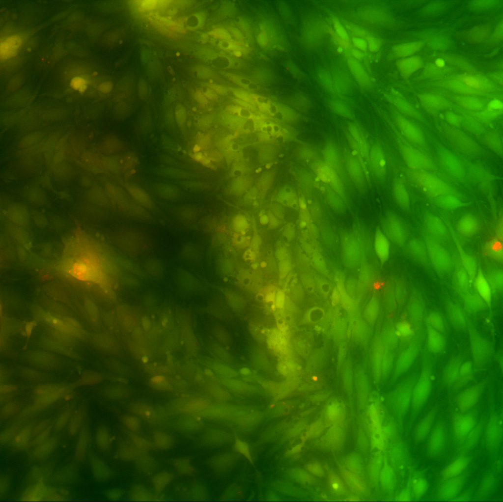 Unfortunately, the TMRIA also non-specifically labeled the cells – notice the yellow overlay throughout these images. This particular rhodamine conjugate is thiol-reactive so that’s not surprising. However, none of the ‘vacuoles’ fluoresce red, as was expected. Assuming that some of the dye remained unconjugated (to thiols), some could have been pinocytosed (but wasn’t). If it’s worth pursuing these experiments further, I need to optimize the Dextrans protocol or get a non-reactive carboxytetramethylrhodamine.
In the last image there appears to be red concentrated in the few ‘vacuoles’ I can find over the supported areas. Do you agree?
The yellow spot toward the bottom of the membrane is not a vacuole – it’s stained both bright green and red separately. The red blotch furthest to the right might be a vacuole – but it’s on supported pnc-Si. The intermediate red spot is not a vacuole – I can show the red and green channels separately if you want.