Microscope Scale Electrophoresis
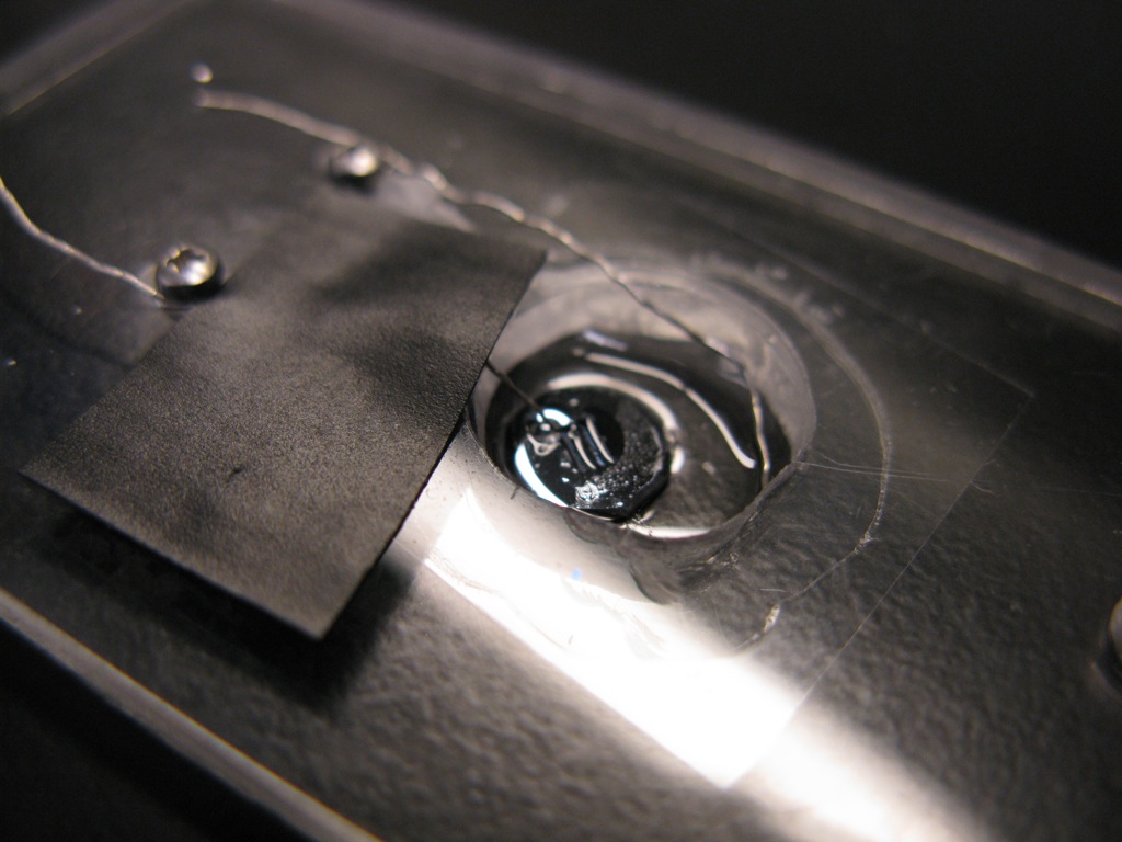
Today I built a small scale electrophoretic cell using the components from Lingyun’s study. I drilled out his cell and affixed a coverslip to the bottom. I set up a pinholed membrane just like our standard diffusion assay (with silica beads and PBS underneath the membrane). Two platinum wires were used as electrodes with the negative electrode in the PBS reservoir and the positive electrode touching the silicon support near the wells (trying to get the wire actually in the well resulted in broken membranes).
I then pipetted 5ul of .5mM rhodamine on top of the well so that the electrode was immersed. I turned the power supply on to 10V (~3mA according to the supply). I took images every .2 min for 20 frames. Half way through the experiment I switched the polarity of the electrodes. It appears that the rhodamine front moves back toward the slit, but as time passes it also looks like the rhodamine is being quenched. There was very little fluorescence in the entire cell at the end of the experiment.
Here is a movie:
I was not observing electro-osmosis during this experiment.
On a separate note, I showed yesterday that there is little or no observable passage of water through an intact membrane up to 60V for 40min. This same membrane was able to pass PBS and rhodamine through to the negative electrode. This is important because it is the first time we have seen a separation using electrophoresis in an intact membrane.