Fluorescence Adsorption Assays
Glass chips and pnc-Si membranes were incubated in 1mg/mL FITC labeled BSA for 30min. They were vigorously washed in a large beaker of PBS. Chips were placed on a slide and imaged with fluorescence scope.
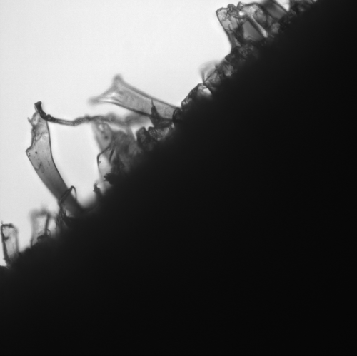
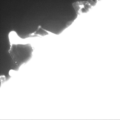
For this study, it seems that the fluid surrounding the chip was much brighter than the chip itself. The following cross section movie pans across the membrane.
DIC image on top, FITC image on bottom
I will be preparing the chips and drying them after non-adsorbed BSA is rinsed off. This might give us a better representation of the actual adsorption to the chip.
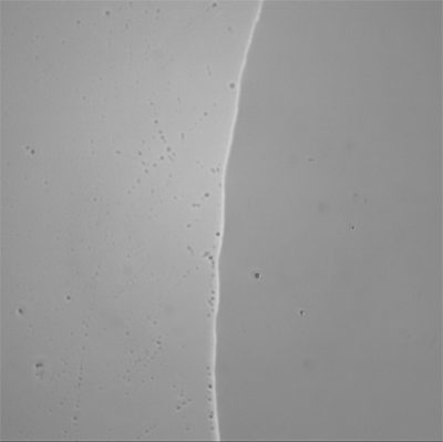
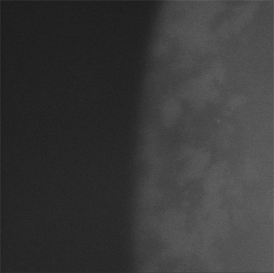
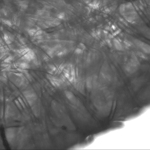
I additionally took some images of Millipore cellulose and PES ultrafiltration membranes. We have said before that the membranes are autofluorescent and thus can not be used for this adsorption assay. Indeed these next images (which are thresholded similar to the pnc-Si fluorecence images) show that the polymer membranes are too fluorescent to perform this assay.
Cellulose Membranes (DIC and FITC images)
PES Membranes (DIC and FITC)