HUVEC Tight Junction and Cadherin Immunofluorescence
In my last post on HUVEC intercellular junctions, I just showed cadherin staining. I repeated this protocol but also tried immunofluorescence for ZO-1, the tight junction protein.
These are HUVEC P7, 6 days post-seeding @ 50,000 cells/cm2. I looked at glass coverslips, pnc-Si and commercial PET transwells. Since background fluorescence was so high in my previous images, I used the reducing agent sodium borohydride to try to decrease background. I also used different antibody dilutions to see if there was a more optimal concentration for staining (1:500, 1:500 for cadherin, 1:40, 1:100 for ZO-1). I also counterstained the nuclei with Hoechst 33342 (H33342).
Glass coverslip treated with sodium borohydride stained for cadherin (H33342 counterstain)

Glass coverslip treated with sodium borohydride stained for ZO-1 (H33342 counterstain):

There was clearly no staining of intercellular junctions (either cadherin or ZO-1) in either of these slides. It looks like the background went down (at least for TRITC red) compared to my earlier post, so sodium borohydride seems to help.
pnc-Si stained for cadherin (H33342 counterstain):
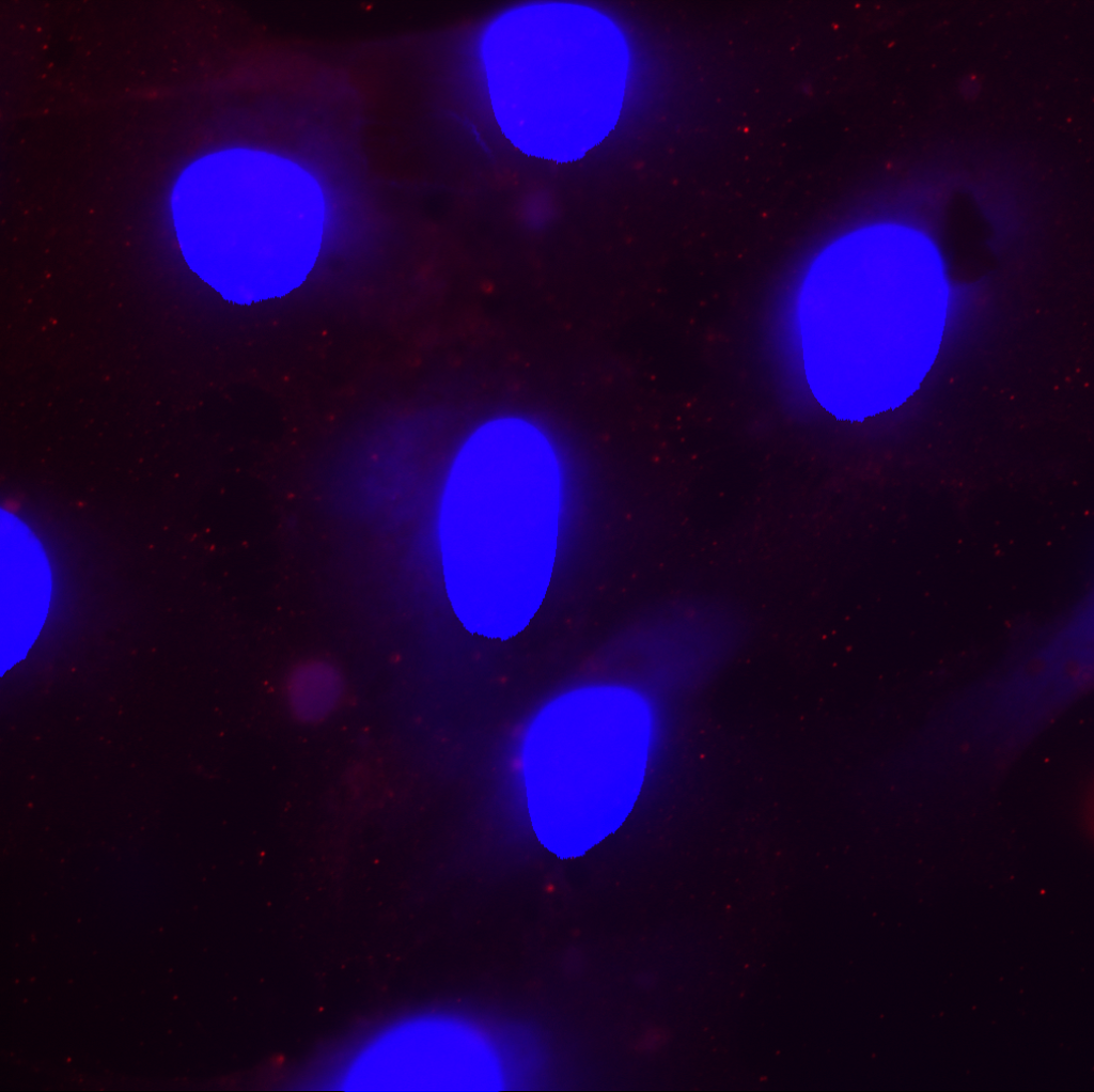
Here. the background is really low compared to my earlier post. This indicates that pnc-Si is not a source of extreme autofluorescence in red or blue channels. Unfortunately, the staining was punctate and there was no evidence of cadherins.
PET transwell sequentially stained for cadherin and ZO-1 (H33342 counterstain):

Here I tried to be fancy and stain for cadherin (red) and ZO-1 (green) at the same time, with H33342 for the nucleus. Again, no intercellular junctions are obvious. What is remarkable about this image is the extreme autofluorescence of the PET membrane. It’s so bright in the green channel that it almost overpowers the background cell staining by this antibody.
This experiment was largely disappointing since no intercellular junctions were observed. It’s possible that the cells were a little too old (P7) to express these proteins. It’s also possible that my antibody titrations were not ideal for HUVEC, and I didn’t have enough antibody to properly stain these proteins. I don’t think the problem is with sodium borohydride since there was no staining in non-treated pnc-Si and PET samples. I am moving on to doing Ab titrations with the new bEnd3 cell line.