f-BSA adsorption on Functionalized pnc-Si (Shestopalov Lab)
Katia in Dr. Shestopalov’s Lab functionalized some pnc-Si windowless chips. The process she follows is shown in Figure 1. She provided chips at various stages of the process. “Grignard”, “Diazirine”, and “PEG”.

After functionalization, the chips were wetted with 20 µL of 1:1 pbs then 40 µL of 5 mg/mL f-BSA was added. The chips were incubated for two hours in a refrigerated petri dish (sealed with DI water soaked Kim wipe). The chips were rinsed with pbs and DI water the dried with nitrogen. In addition to the functionalized (and partially functionalized) chips, a bare pnc-Si chip was also incubated with f-BSA.
The fluorescence ‘scope was used to capture images and ImageJ used to measure the intensity. The results of the measurements are shown in Figure 2.
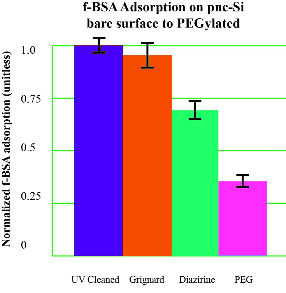
The results in Figure 2 are without the background signal removed. (this is any fluorescence recorded from the pnc-Si surface or the PEG layers.
The measurements show no difference between the bare chip and the “Grignard” chip. There is a progression of improvement at the penultimate “Diazirine” step to the final PEGylation step.
Figure 3 shows the normalized fluorescence from a bare pnc-Si chip and a chip with a functionalized surface, no contact with f-BSA.

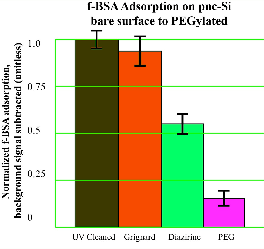
Subtracting the background signal of the PEGylated pnc-Si leads to the results shown in Figure 4. This shows a reduction of the adsorption of f-BSA to ~15% of that on bare pnc-Si.