Adipose Derived Stem Cell Differentiation – Recap
Over the weekend, I resubmitted our adult Adipose-Derived Stem Cell (ADSC) paper titled:
Membranes Promote Endothelial Differentiation of Adipose-Derived Stem Cells and Perivascular Interactions
There were three main findings:
1.) ADSCs can be induced to express CD31 (PECAM-1) like endothelial cells.
a.) Several prior studies used Vascular Endothelial Growth Factor (VEGF) at 50 ng/mL to induce expression of CD31. When we used this approach we found that the cells began to express CD31 (ADSCs are initially tested to be negative by flow cytometry), but the expression was uniform and not a typical endothelial cell (EC) distribution at cell-cell borders (see the first two pictures of this post). This non-EC expression was also seen by some of the other groups, but they still reported it as a positive finding.
b.) One prior study used reduced serum media without any growth factors. We saw minimal increase in CD31, which might not have been significantly different from cells cultured in proliferation media.
c.) When we co-cultured ADSCs with HUVECs either directly across the membrane or separated by 900 microns, we found that a subset of the ADSCs expressed CD31 at cell-cell borders like confluent ECs. We never found all of the cells to do this, but this is not uncommon in the stem cell world. Many differentiation protocols are considered successful if yields are between 10-50%. 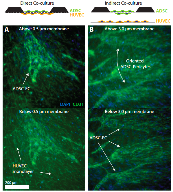 .
.
2.) ADSCs also act like pericytes when cultured with ECs.
There is a large body of literature and some controversy over whether mesenchymal stem cells are in fact pericytes or whether MSCs/pericytes evolved separately but have similar functionality.
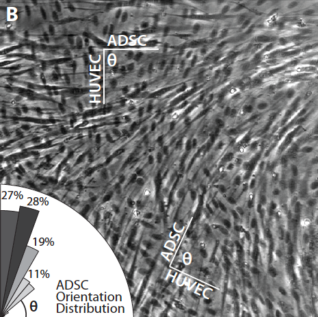
A.) We have found in a few instances that ADSCs will orient perpendicularly to polarized HUVECs across a 0.5 micron low porosity membrane. I qualified HUVECs as polarized if their nucleus had as aspect ratio >1.5:1 and then measured the angle between the cells in three different images. The mean angle was 68 +/- 16 degrees (n=264). Representative image and rose diagram is shown below.
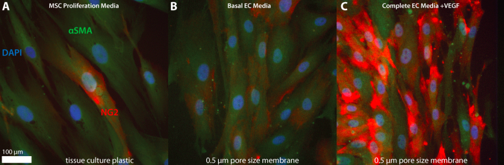
B.) Others have defined several pericyte markers. One of these markers is alpha smooth muscle actin, which is obviously found in smooth muscle cells, but also pericytes (mural cells). Another marker is NG2, which stands for neural/glial antigen-2. NG2 is found on MSCs, but not necessary at high levels. After exposing ADSCs to either proliferation media, basal media (w/o growth factors) or complete EC media plus VEGF, we found interesting results. The ADSC population in PM showed a very heterogeneous expression of NG2, with all cells expressing SMA. In basal media on 0.5 micron membranes the number of cells with NG2 may have increased, but the expression levels were unchanged. In complete EC media with VEGF, NG2 levels were 2-3x higher, with the majority of cells expressing NG2. In the image below, exposure and intensity min/max levels are identical between the images (all collected on the same day with the same original flask of ADSCs, P5).
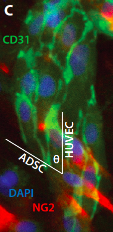
During ADSC-HUVEC direct co-culture, I also stained for NG2. Not surprisingly, the ADSCs that oriented stained strongly for NG2. Interestingly, they did not seem to express high levels of CD31.
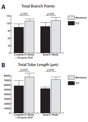
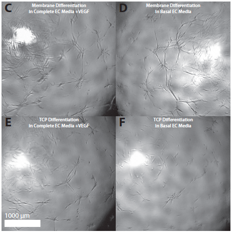
3.) ADSCs induced in basal or complete EC media have significantly greater angiogenic potential in a standard angiogenesis assay if they are induced for 6 days on a membrane versus on TCP. The analysis of the angiogenesis assay was performed at 18 hours after seeding.
Representative Images:
Conclusion – Membranes and co-culture with HUVEC help ADSCs differentiate into a range of vascular cells. Future work will need to focus on:
1.) What improved procedures/protocols will commit a higher % of ADSCs to one lineage or the other
2.) If it’s not feasible to commit entirely to one cell type, is it possible to separate the cells after differentiation?
3.) Will the mixture of differentiated cells sort everything out on their own in vitro or in vivo? Is it necessary to deal with individual cells types?
4.) Are we also creating or can we create smooth muscle cells?
5.) All of these induction experiments were only conducted for 6 days, what happens if we go for 14-21 days? How do we deal with over-confluence?