Manual Reconstruction of Electron Tomograms
Using the techniques described in my previous post: TEM Tomography of NPN and npMgF2, we have recreated some tomograms that have contour (position) and height (depth) information contained in them. Our goal now is to express that height information in the form of a surface, or volume.

Segmentation
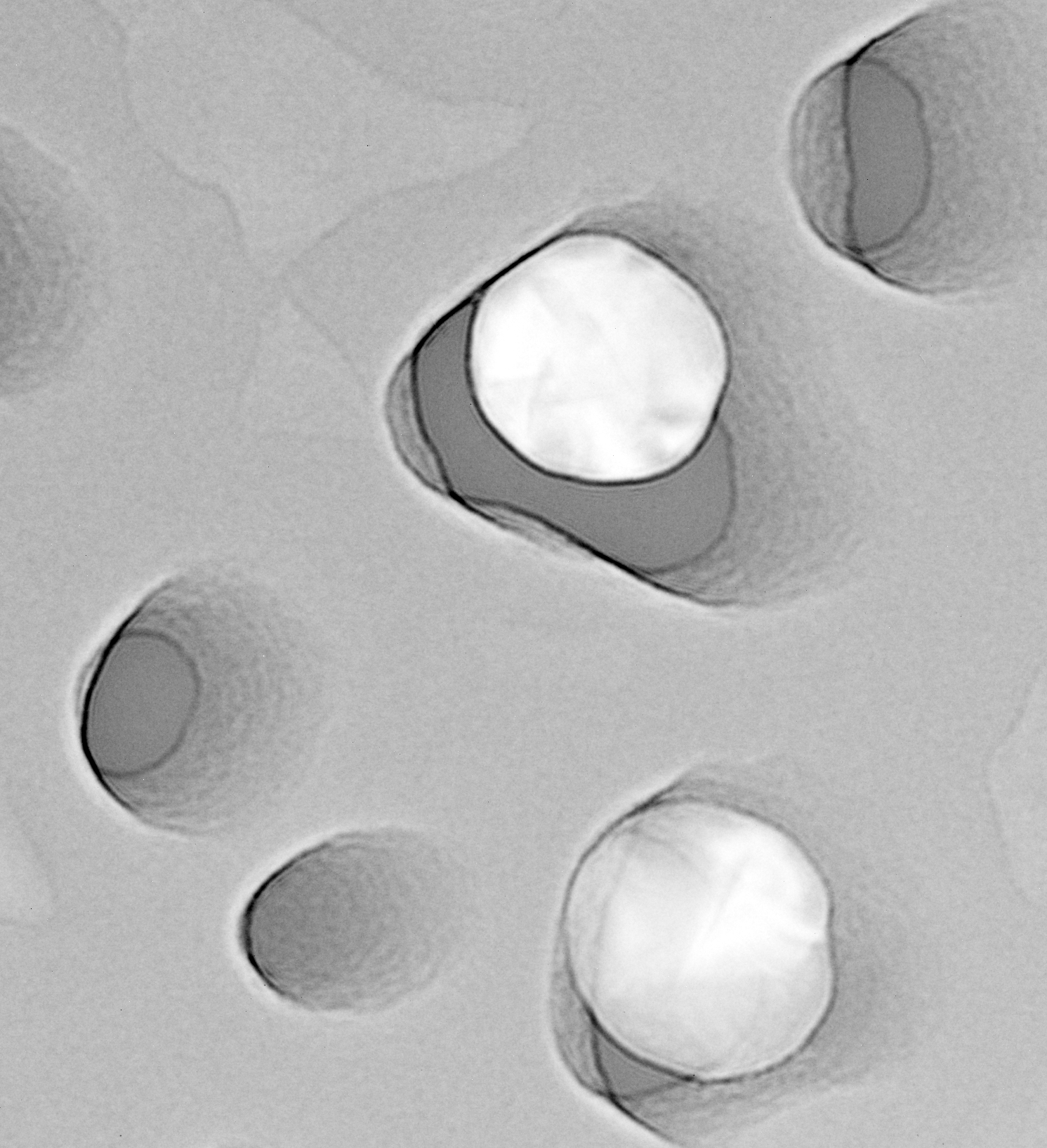
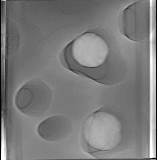
Having some evidence that our reconstruction method is valid, we can proceed to highlight different contours in the tomogram stack. By tracing the interesting contours individually, we can rebuild an interpolated mesh throughout the layer. The precision of the recreated mesh depends on the number of slices in the tomogram and the contrast that allows us to identify the contour. While there are automatic methods to segment structures available, I have not identified the best procedure to reproduce accurate features.
Curves were traced from each slice of the tomogram stack. The red cones are the inner contours of the NPN pores. The Au nanoparticle (yellow, ~60 nm spheres) positioning is based on inference in the tomogram; it is so electron dense that the sphericity/positioning is lost. The orange sheets are the siloxynitride scum on the surface of the NPN. Each hole tapers to the bottom of the film, except for one which is closed off. Thus far, I have manually recreated contours by tracing them every so often down the height stack (4 or 5 contours). A more precise recreation requires tracing every slice, and this will be required for the much less homogeneous MgF2 volcano structures.
Volume Representation
FreeD also permits these structures to be exported in an STL format for easy 3D printing. With a little blender magic, I can render these contours in a block.


Future Improvements
- Automatic segmentation would obviate the need for tracing these contours by hand, and eliminate human bias. We’re still relying on a little inference for tracing these contours.
- Tomograms can be improved by
- increasing the resolution of the tilt stacks (1 deg increments instead of 3 deg increments). This will lessen the ‘ringing’ behavior in the tomogram.
- increasing the range of the tilt stack (the full 60 deg offered by the stage instead of only 30 deg total). We will get more precise positioning information in the tomogram.
- better tomogram production using more advanced algorithms