Au Nanoparticles deposited on tented structures
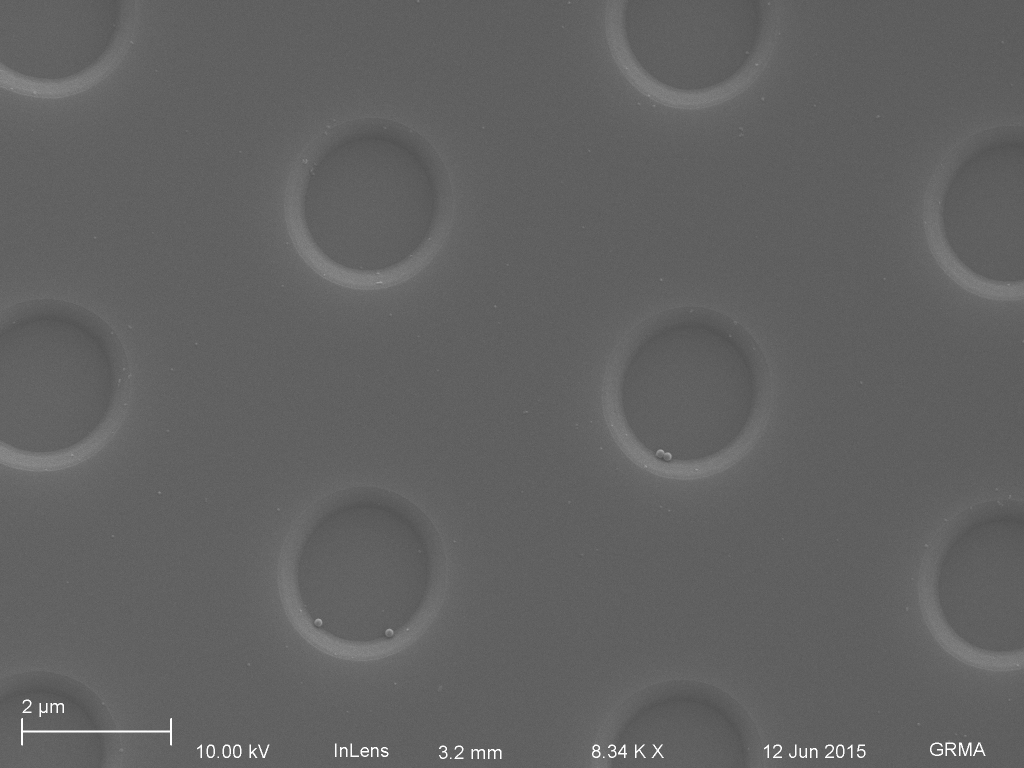
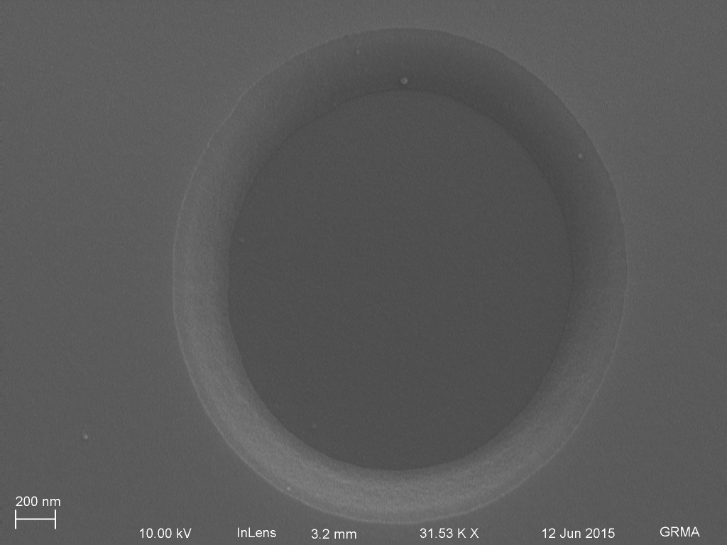
To test the filtration capabilities of our tents, Tucker and I immersed a tented (wafer 1150, ~30 nm pores) oxide structure (~3 um hole diameter, 200 nm high) and a nontented structure in a solution containing 60 nm (.043 nM) and 10 nm (~10 uM) Au nanoparticles for 1 hour (2 uL incubation volume), rinsed them (100 uL), dried them out (70C oven 1 hr), and imaged the remaining particles and structures under SEM. Unfortunately, there were no particles remaining. Experience from Josh Winans has told us that gold generally does not like to attach to silicon nitride.
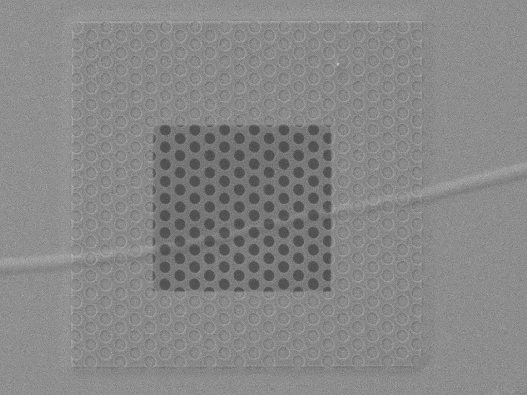
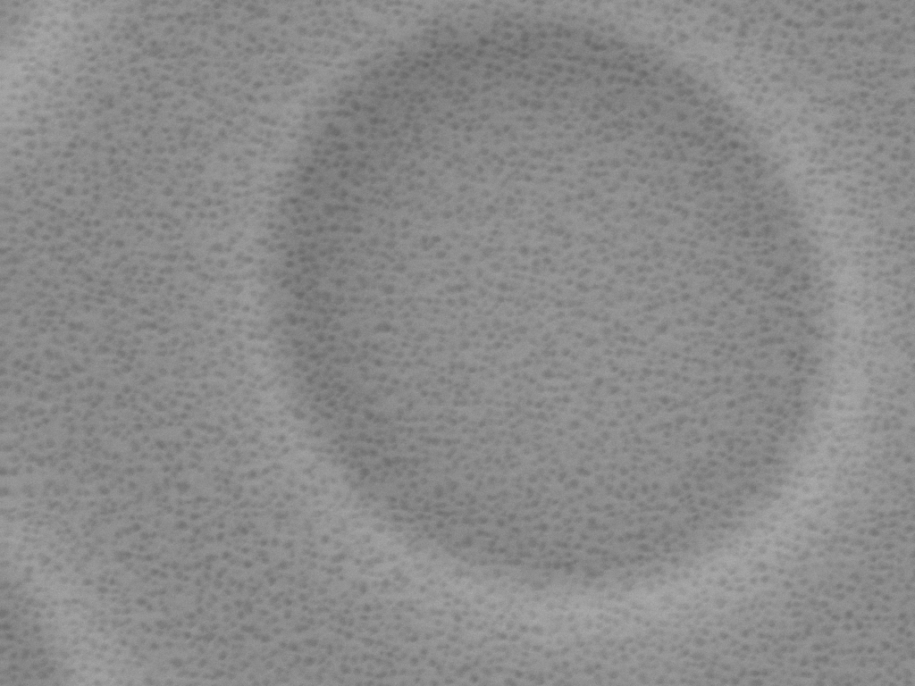
So we switched from gold particles to polystyrene beads (20 nm and 100 nm diameter). These substrates were coated with 5 nm platinum before imaging. Unfortunately, the tent flew off the active region, but the images are still useful.


We still need to show a good crossection of this process. Two images would be sufficient.
1. An image like ‘Tent acts as a prefilter’, displaying the nanomembrane filtering out garbage from the cavity.
2. Fractured membrane with small particles below, and no large particles below.
Using a tent chip substrate with posts instead of holes would increase the chances of finding picture number 2, as it increases the area below the tent. We’ve also observed that the polystyrene tended to associate with the oxide spacer instead of the nitride floor in the attocavity space. Moving to a substrate with posts will help us by reducing the amount of oxide overall compared to the pit substrates.