Fibroblast wound healing with small EVs
Wound healing experiments
I have been isolating extracellular vesicles (EVs) from adipose-derived stem cells (ADSCs) and using them in wound healing experiments. For isolation, I follow the same protocol as outlined here. For these experiments, I used small EVs, which are collected in the last step of the isolation process.
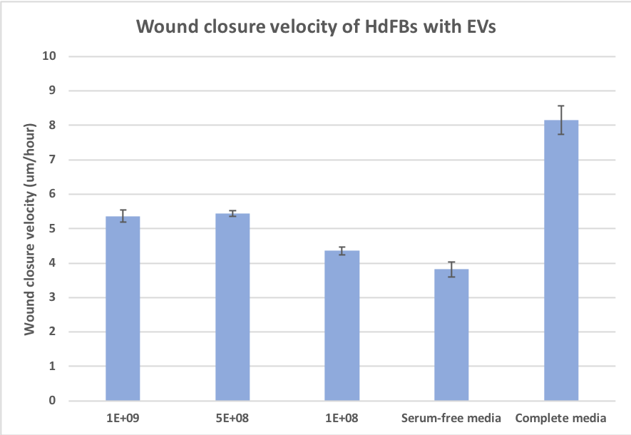
I began by seeding human dermal fibroblasts (hDFBs) in a 24-well plate. Cells grew until they reached 90% confluence. I then created a wound in the bottom of each well using a p200 pipette tip. Experimental wells were given serum-free media (DMEM) supplemented with different concentrations of isolated small EVs (1E9, 5E8, or 1E8 EVs/mL). Other wells were treated with either serum-free media or complete media (DMEM + 10% FBS + 1% P/S), acting as the negative and positive controls. Using the MATLAB tracking program created by Henry, I obtained the wound closure velocity of each condition over the span of 48 hours.
The results shown below come from two separate wound healing experiments. As expected, none of the concentrations perform as well as the positive control. These cells prefer complete media with FBS over ADSC EVs. However, all three EV conditions heal the wound faster than the negative control. This could indicate that ADSC EVs impact the wound healing ability of hDFBs.
EV counts and ratios
One goal for this project is to determine the physiological relevance of the wound healing experiments performed by myself and others. Typically, people dose with large concentrations of EVs. While this showcases the effect of EVs on a large scale (possibly for a drug-related application), it does not provide insight into their effects on a physiological level. To be more conscious of this in my own experiments, I have performed a few calculations (see explanation and math below).
Using NTA, I have measured that ADSCs (total number of cells = 677,700 +/- 1,417) create roughly 5E9 EVs over the course of 24 hours. This is 7,378 EVs produced per ADSC every 24 hours.
In my experiments, I worked with three different concentrations of EVs: 1E9, 5E8, and 1E8 EVs/mL. Each well contains 800 uL of media. Therefore, there are either 8E8, 4E8, or 8E7 EVs present in each individual well, respectively. Using a MATLAB cell counting program, I have also determined that there are roughly 85,000 hDFBs in each well. This results in a total of 9444, 4722, or 944 EVs present per hDFB.
By dividing the number of EVs present per hDFB by the number of EVs produced per ADSC, I am left with a new ratio. It represents the number of ADSCs needed to produce EVs per hDFB. For example, the highest concentration that I tested (1E9 EVs/mL) has a ratio of about 1.3:1 ADSC:hDFB. This means that for each hDFB cell, I would need 1.3 ADSCs producing EVs to reach the desired concentration. For my three tested concentrations, the number of ADSCs needed for each hDFB is 1.3, 0.64, and 0.16. From these calculations, I conclude that my concentrations are already (at least somewhat) relevant to physiology, as my highest concentration is nearly 1:1. I suspect that other groups who are conducting similar work have ratios much higher than mine, though I have not yet performed additional calculations.
Math
Amount of EVs produced by ADSCs
Amount of EVs in 2.14.20 stock: ~5E+9 EVs
5E+9 EVs/677700 cells = 7378 EVs produced by each ADSC
Amount of ADSC EVs present in each well of the 24-well plate, depending on condition
1E+9: 1E+9 EVs/mL * 0.8 mL per well = 8E+8 EVs
5E+8: 5E+8 EVs/mL * 0.8 mL per well = 4E+8 EVs
1E+8: 1E+8 EVs/mL * 0.8 mL per well = 8E+7 EVs
Amount of EVs present per hDFB in the wound healing experiment, depending on condition
1E+9: 8E+8 EVs/84708 HdFBs = 9444 EVs/cell
5E+8: 4E+8 EVs/ 84708 HdFBs = 4722 EVs/cell
1E+8: 8E+7 EVs/84708 HdFBs = 944 EVs/cell
Ratio of ADSCs to hDFBs
1E+9: (9444 EVs/HdFB)/(7378 EVs produced/ADSC) = EVs from ~1.3 ADSCs (over 24 hours) per FB in the 24-well
5E+8: (4722 EVs/HdFB)/(7378 EVs produced/ADSC) = EVs from ~0.64 ADSCs (over 24 hours) per FB in the 24-well
1E+8: (1180 EVs/HdFB)/(7378 EVs produced/ADSC) = EVs from ~0.16 ADSCs (over 24 hours) per FB in the 24-well
Interesting post. Is 1:1 ratio determined as the ratio of the ADSCs to HdFBs in your target tissue based on literature?
How many repeats you had in each separate experiments?
As of right now, the 1:1 ratio is an estimation. I have yet to find the true ratio in literature, though I plan to do so.
I had two repeats for the wound healing experiment. Each condition contains multiple regions of interest from each experiment. The math was conducted using a few different cell images and rough averages of the two experiments I explained in the beginning of the post.
Comment to myself/Hayley – We need to still do the calculation to determine how much of original conditioned media makes into the final EV dosing to determine the factor reduction in growth factors excreted by the ADSCs into the conditioned media. I think it was several orders of magnitude based on the serial centrifugation steps, but can’t remember. Alternatively and probably even better would be to run an ELISA for VEGF and/or FGF in the conditioned and dosed media. Downside to the ELISA is that we have to pick particular growth factors to test.
Related comment – I forgot why we dropped the condition where we use serum, but “EV deplete” it via ultracentrifugation.