Membranes on STEM
Chris and I got to play on the new Zeiss scanning transmission electron microscope (STEM) today (learn about STEM here). We didn’t get to spend a lot of time on it, but here are some quick images we snapped.
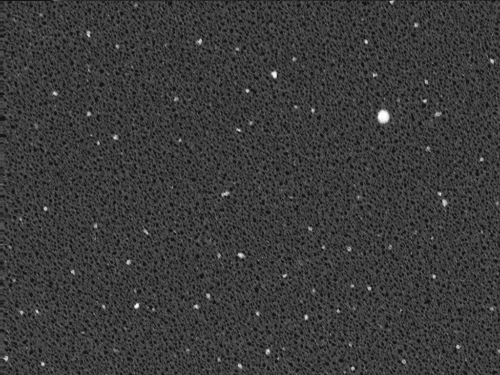
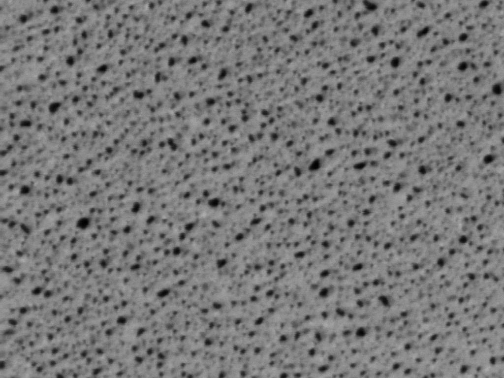

wafer 416 @ 25kx (left) and 65kx (right)
Even though we’re imaging in transmission mode, the pores appear dark because we are using the dark field detectors (picking up off axis electrons). One difficulty we had was that imaging in one spot for an extended period of time deposited a film (maybe carbon) that occludes the pores. We’ve also noticed this using the URMC Hitachi TEM. Whatever contaminant we’re introducing inside the chamber is showing up as bright spots in the dark field image (see 25kx image).
We should be able to clean the images up a bit once we have more time on the tool to tweak some of the settings.