Gap Junction Formation
Background: The purpose of these experiments was to investigate the formation of gap junctions between co-cultured HUVEC and ADSC across 0.5µm HP SiO2 and 0.4 µm HP Track-Etched membranes. Gap junctions are channels that allow small molecules to pass directly from cell to cell. Electron micrographs show that the membranes of two adjacent cells are separated by a uniform gap of 2-4 nm. The gap junction channel, the connexon, is synthesized by specialized channel-formin proteins, connexins. We hypothesized that gap junctions will not form between the cell layers cultured on Track-Etched membranes because the membrane is too thick (~10 µm). Our primary investigation was to determine if the SiO2 membranes were thin enough (~300 nm) to allow the formation of gap junctions.
Methods:
- Create cell culture plate, bake in oven at 70-80C overnight, and UV sterilize both sides for 15 minutes
- Coat both sides of the membranes with 1% Geltrex, incubate at RT for 30 minutes, and aspirate solution
- Seed HUVEC on the bottom of each membrane at a cell density of 5000 cells/cm²
- When HUVEC reach confluency (2-3 days), stain cells with 1µM CFDA
- Seed ADSC on top of each membrane at a cell density of 5000 cells/cm²
- After 2-3 days, stain ADSC with 1:200 anti-NG2
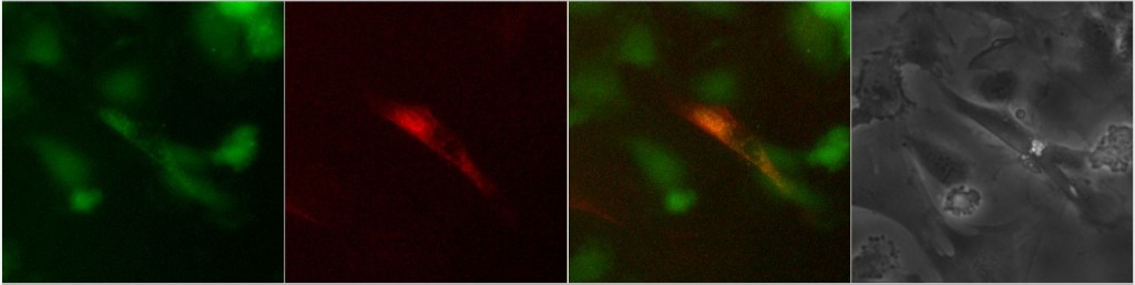
- Image on widefield microscope using GFP and TxRed filter cubes and phase contrast
Results:
ADSC stained both green (CFDA) and red (anti-NG2) would appear yellowish in the overlaid fluorescence images, and would indicate formation of a gap junction. Several trials were executed and evidence of gap junction formation on 0.5 µm HP SiO2 only occurred once.