Intact pnc-Si with HUVECs after 1 week
My last cell post involved pictures of HUVECs after 1 day of growth on commercial transwells and pnc-Si Sepcons. Over the past week, I’ve been growing HUVEC on commercial PC transwells and pnc-Si in the Sepcon format in order to track the TEER. To get more data out of these cells, I decided to do some microscopy. All of these images are from day 7.
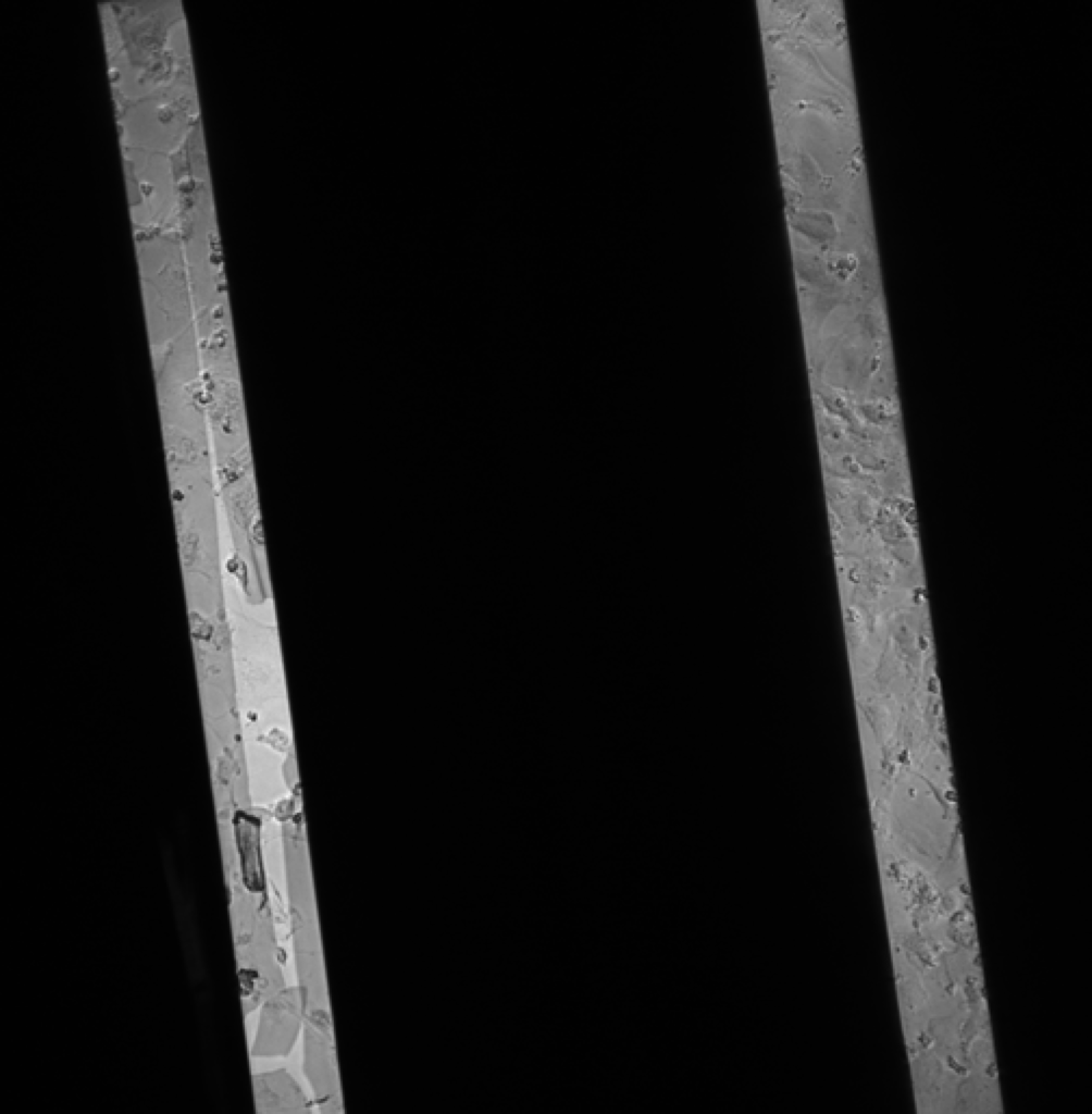
This is a 10x phase contrast image – the left membrane is clearly broken. However, the right membrane slit is intact, with what appears to be a HUVEC multilayer on it. The left membrane was actually intact before I started the fluorescent staining protocol. Remember that this is after 7 days of HUVEC growth, so the membranes were intact after ONE WEEK of cell culture in the Sepcon format!!!!
This image is from a different pnc-Si Sepcon.
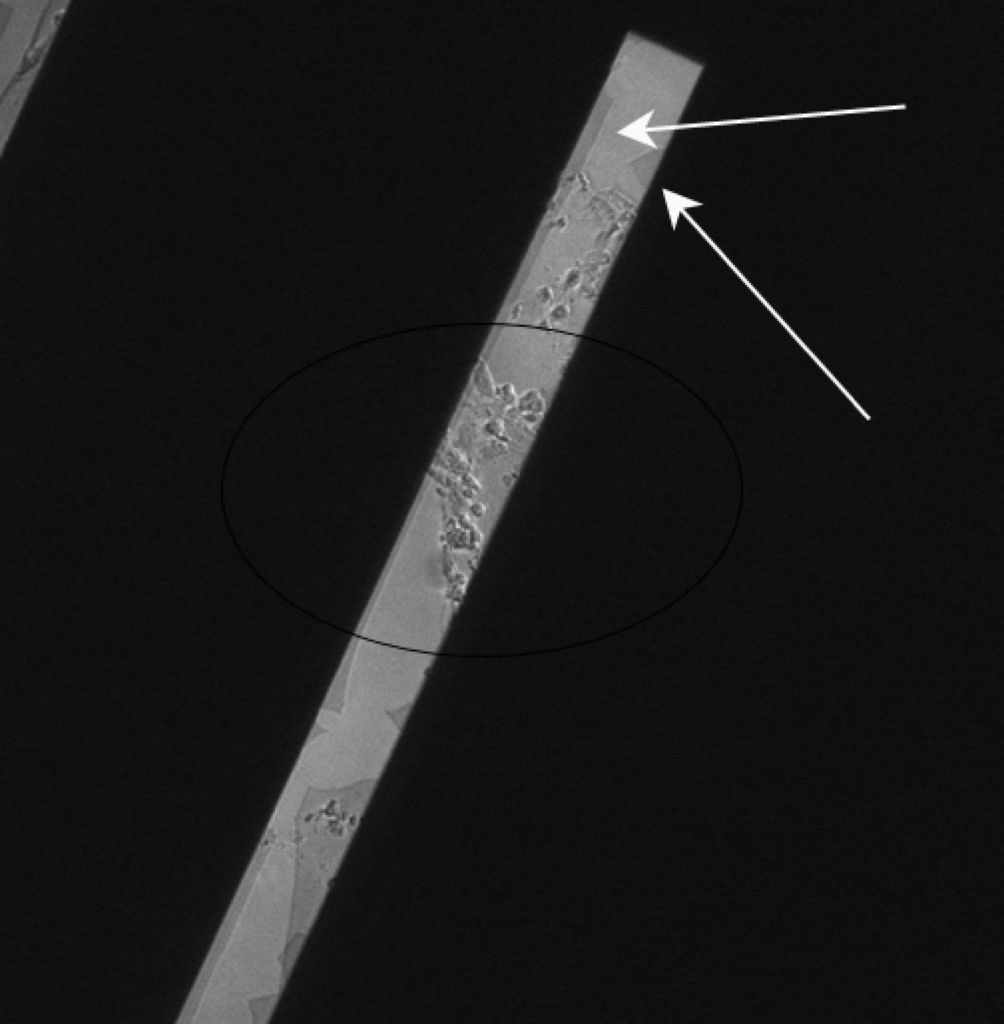
I included this picture since it shows HUVEC growth across a membrane slit even without the membrane present. The white arrows point to the broken membrane; if you follow the membrane edge, you see that the slit is entirely broken. The black circle highlights an area of HUVECs that are across the slit, basically hanging in free space.
Was there anything special about this sample? Is this the type of madia that would typically dissolve the membrane in 24 hours? Do you think it’s the cells that are promoting chemical stability? Do they consume bicarb or produce H+?
Nope, nothing special that I can think of. It’s regular cell media for HUVEC (EGM WITH FBS). Since this wasn’t a confluent monolayer, I doubt that cells are blocking media access from the membrane side. I think cells consume some bicarb. Based on my experience, cells tend to acidify media over time (turn phenol red yellowish). I imagine that all types of microenvironments could be formed between cells and the membrane, though.