Epithelium on transwells(2)
My last post was the first experiment I performed with rat lung epithelial cells on transwells. It seemed like there was vacuolization but it was delayed compared to endothelial cells (and only happened on 1 of 2 pnc-Si transwells). Here, I did a repeat with a lower seeding density (25000 cells/cm2) and with SC613.
The results after 1 day are similar to the first experiment – few/no vacuoles on PET or pnc-Si. The density is lower and there is less of a multi-layer morphology because the seeding density was lower.

After 3 days, there are small and scattered vacuoles on PET. There are also vacuoles on both samples of pnc-Si, although they are not as smooth, well-defined, large or as numerous as for endothelial cells. Epithelial cells on supported pnc-Si also expressed vacuoles (right-most panel).
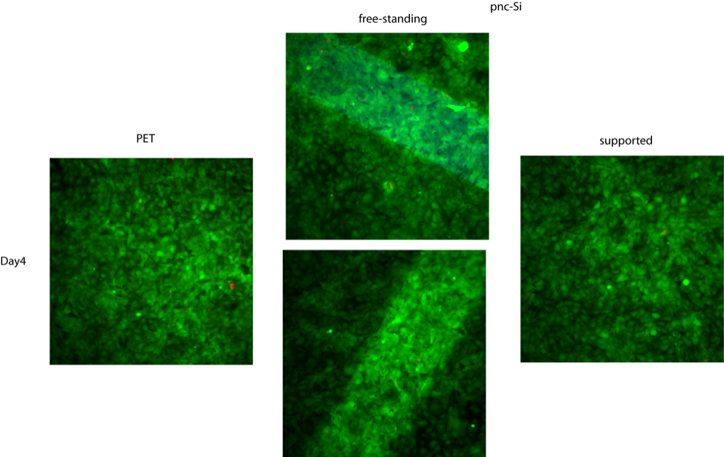
So, this experiment also shows a time-dependence for vacuole formation in epithelial cells. However, vacuolization is not as dramatically limited to free-standing pnc-Si as it is for endothelial cells. The morphology and density of the vacuoles is also different than for endothelial cells.
I’m not sure where this leaves me. We set out to show that vacuolization is endothelial cell specific, which these results contradict. However, epithelial cells also form tubes in vivo and in vitro but I haven’t read many papers about epithelial tubulogenesis. Therefore, I’m not sure if I should include this in the paper to demonstrate the generality of vacuole formation in lines of cells that can form tubes, or if that would instigate too many questions.
If you can so no vacuoles for fibroblasts that look reasonably confluent, lets include that.