hiPSC Differentiation into Neural Crest Stem Cells
Introduction
Breakdown of the blood-brain barrier (BBB) is one of the earliest signs of sepsis and is linked with long-term cognitive impairment. As animal models poorly recapitulate human sepsis, alternative approaches are direly needed to understand sepsis-associated brain injury and develop strategies that protect against cognitive decline. To fill this gap, we are developing a microfluidic model of the human blood-brain barrier (µSiM-hBBB) to elucidate mechanisms of BBB breakdown during sepsis. The first version of the µSiM-hBBB will be developed using human induced pluripotent stem cells (hiPSC) to derive two cellular components of the neurovascular unit: brain microvascular endothelial cells (BMECs) and pericytes. While BMECs are the main component of the BBB, pericytes are key support cell of the neurovascular unit (NVU), along with astrocytes and microglia. Pericytes not only help stabilize the BBB, but are key responders during inflammation and help guide immune cell transmigration.
Several protocols have been developed to differentiate pericytes from hiPSCs, however, only recently, a protocol was established to develop brain pericytes following the cells’ developmental pattern (Stebbins et al., Neuroscience 2019). This method drives hiPSCs towards neural crest stem cells (NCSC, p75-NGFR+HNK-1+) and then guides them into NG2+PDGFRβ+ “brain pericyte-like cells”. This post summarizes my first attempt at replicating this protocol in our lab, focusing on differentiation into NCSC.
Methods
Thaw Cells (WiCell iPSC (IMR90)-4)
- Thaw iPSCs briefly in a 37°C waterbath, swirling gently.
- Transfer gently to 15 mL conical and add 11 mL Essential Medium 8 (E8, gibco) with 10 µM ROCK inhibitor (Y-27632, Millipore) dropwise to cells, gently swirling as media is added.
- Pellet cells by centrifugation at 200 x g for 5 min and resuspend in 1 mL E8 + 10 µM ROCK inhibitor.
- Add 500 µl/ well cell suspension to 1.5 mL in Matrigel-coated 6 well dish. Incubate at 37°C, 5% CO2.
- Feed cells with E8 medium daily. Pass when cells reach 70% confluency.
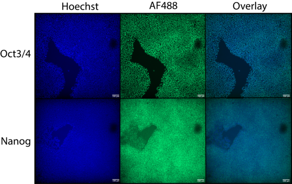
Plate a subset of cells for ICC analysis to confirm pluripotency (Oct 3/4 and Nanog)
NCSC Differentiation
D-1
- Singularize cells with accutase and add to 4 times DMEM/F12 (1X) (gibco).
- Pellet cells by centrifugation at 200 x g for 5 min and resuspend in E8 + 10 µM ROCK inhibitor to a density of 8.75 × 10⁵ cells/well and add to Matrigel-coated 6 well plate.
- Incubate at 37°C, 5% CO2 for 24 hours.
D0
- The following day, switch medium to E6-CSFD: Essential Medium 6 (E6, gibco) supplemented with 22.5 µg/ml heparin (Millipore), 1 µM CHIR99021 (Millipore), 10 µM SB431542 (Tocris), and 1 µM dorsomorphin (Millipore).
D1-14
- Feed cells with E6-CSFD daily. Pass when cells reach 100% confluency.
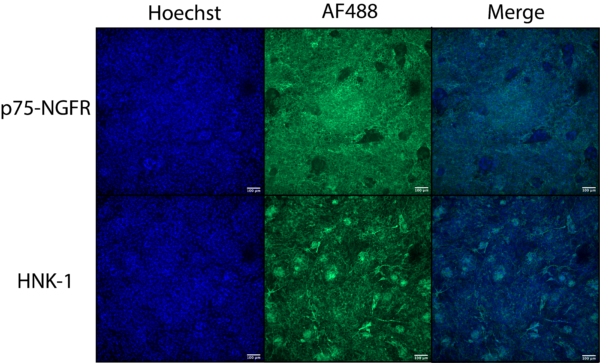
Plate a subset of cells for D15 ICC analysis to visualize NCSC phenotype (p75-NGFR and HNK-1)
Purification of p75-NGFR+ NCSC
D15
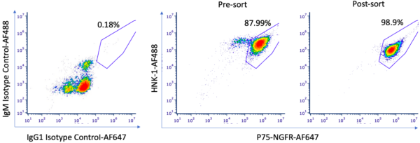
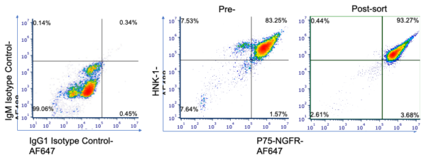
- Sort p75-NGFR+ NCSC via magnetic-activated cell sorting (MACS, equipment and reagents from Miltenyi). Analyze by flow cytometry to determine sorting efficiency.
- After sorting, pellet the eluate at 180 x g, 4°C for 5 min and resuspend in 1 mL E6-CSFD.
- Add to E6-CSFD + 10 µM ROCK inhibitor to a density of 5 × 10⁴ cells/mL and add to uncoated 6 well plate.
- Incubate at 37°C, 5% CO2 for 24 hours.
Results
Conclusions
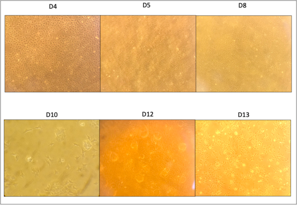
Based on preliminary analysis using ICC, we have successfully differentiated hiPSCs into NCSCs. During the first 15 days of differentiation, cells were guided to NCSCs, with morphology matching Gastfriend et al (near submission?). Importantly, by day 12, we saw clusters of cells that were p75-NGFR–, which were subsequently sorted out via MACS. Flow cytometry analysis technique, while changing the percent of p75-NGFR+/HNK-1+, does not lead a difference in conclusion. We are still >90% double positive, which is the standard we set for “good enough” to move onto pericyte differentiation.