Sampling Method Permeability Assay Optimization
Motivation
We have discussed the optimization of our in situ permeability assay at length. This assay relies on confocal microscopy, which may not be accessible to all users of the µSiM. Therefore, we developed an alternative permeability assay in the µSiM that is an endpoint, sampling assay. The protocol for this assay can be found here: ALine Modular Device: Sampling Method for Small Molecule Permeability. The theoretical underpinnings of this assay has been discussed in a previous post. Briefly, cells are incubated with dye in the top reservoir for one hour. After the hour incubation, dye is removed from the well and a 50 µl reservoir in a pipet tip is inserted into one bottom channel access port. Dye is then drawn out of the channel from the other port and added to a plate reader to measure fluorescence intensity. Please note it is necessary to add hydrophobic tape or use a hydrophobic pen to prevent media from passively pumping from the reservoir to the other port and spreading across the device surface, resulting in loss of dye.
While preliminary data using this approach is inline with anticipated values for both hCMEC/D3 and EECM-BMECs, we discovered that the sampling process with a dry well above the cells causes total cell loss. This was observed by experiments at UR and in Bern. Unfortunately, loss of cells prevents further downstream analysis, such as cell staining, which is a crucial step for our EECM-BMEC analysis. It is necessary to confirm proper tight junction formation and maturation, or can help reduce device number by using these same devices to observe expression of cell adhesion markers after proinflammatory stimulation. As an alternative, I tested two options: (1) Leaving the dye in the well during sampling, and (2) Removing the dye and adding fresh media back into the well prior to sampling. For each experiment, I used uncoated, cell-free devices, I did one 50 µl sample for wash one, and then added a new reservoir and did a second 50 µl sample for wash two. This second wash was to measure the percent dye recovery from a single wash.
Results 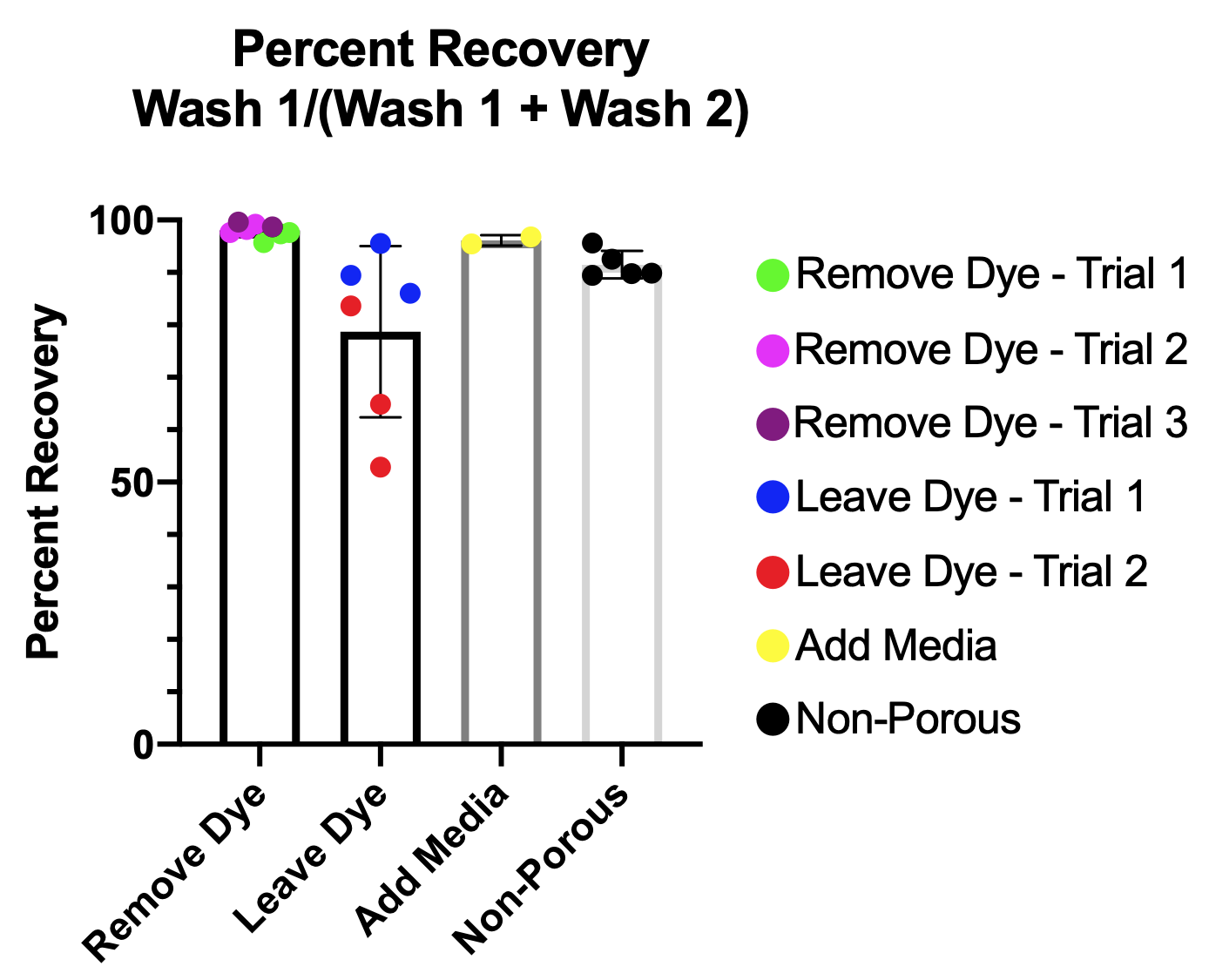
When I remove dye or add blank media back right before sampling, we get ~97% recovery with one 50 µl wash. This is similar to what Howard’s simulations indicate we should see. For the nonporous test, I flooded the channel with a known concentration of dye and sampled 50 µl twice. This resulted in ~90% recovery, which is consistent with previous experiments, and the reason I started using 2 x 25 µl washes. However, taking a second look, sampling dye that is homogeneously mixed throughout the channel is quite different than sampling out dye that is concentrated under the membrane. Given the consistency of the “Remove Dye” data and consistency with Howard’s simulations, I conclude that one 50 µl wash is sufficient to clear dye out of the channel in our permeability experiments. The 3% loss is not enough to significantly change our permeability results.
One striking issue with the “Leave Dye” group is the inconsistency of the data and the poor recovery. The inconsistency is likely due to a splashing issue that occurs when adding the reservoir to one port. Because the top well is full, when I dislodge the pipet from the pipetter, often the movement will cause the fluid in the well to splash. In some cases it would splash onto ports, leading to an erroneously high draw in wash two. Even without splashing, we were likely pulling extra dye through in the second wash due to either lack of cells above or continued diffusion of the dye in the top well into the channel between washes. Both of these issues leading to high dye amounts in the second wash are why we see incorrectly low recovery. The only issue in terms of our real permeability experiments is the splashing that could occur and skew our data.
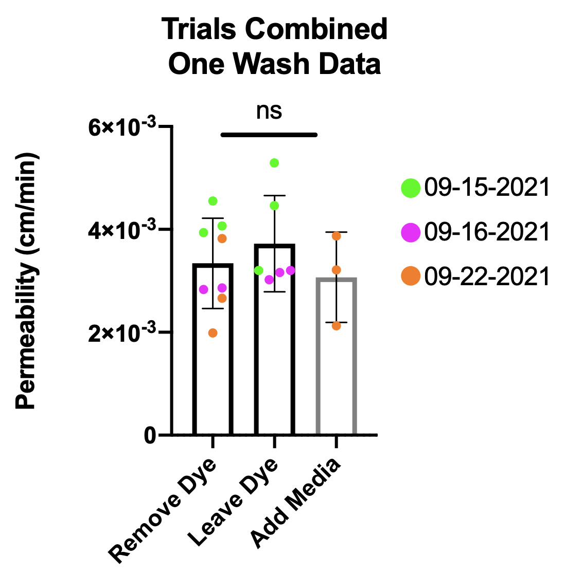
There is no difference in sampling between our three methods, indicating we do not have to worry about continued diffusion skewing our data. In the conclusion, I will discuss the proposed method and order of operations for these experiments.

This is the same data as the two plots previously shown, with each date separate, in case people want to take a closer look. It clearly illustrates that we only need one 50 µl wash.
Conclusion
There is no difference in sampling methods, so we need to pick the method that is best for our cell experiments. We know that sampling on dry cells (“Remove Dye” tests) leads to cell loss. We also discovered that splashing is common when you fluid in the top well during the reservoir addition process. Therefore, the best method is to remove the dye, add the reservoir to one port, and then add media back into the well. Then you can sample from the channel.
I confirmed this process on HUVECs, seeing no cell loss. I then used this method for iPSC experiments and saw data consistent with previous results and was able to stain the cells after the permeability experiment. This was using one 50 µl sample instead of two 25 µl samples. If people agree with these modifications, I will update the protocol shortly.
For those who might do these experiments on several devices:
For the iPSC experiment, since I had 6 cell devices for that experiment (3 NS and 3 TNF/IFN), I staggered the dye addition by ten minutes between groups. This allowed me to sample each group quickly and maintain 1 hr diffusion for all devices. The order went as such for sampling:
- Remove dye all devices for NS group (3 total).
- Then work one device at a time:
- Add reservoir (50 µl)
- Add media well (50 µl)
- Sample channel (50 µl)
- Repeat on next device
- Repeat on TNF/IFN group (I had a couple minutes to spare between groups, indicating 10 minutes was a good timeline).
A second option would be just go one device at a time and note the actual time of dye incubation for each device and use that time when calculating permeability.