Astrocytes on PET and pnc-Si transwells (1)
This is my first attempt to grow astrocytes (NG108-15 cell line) on PET and pnc-Si transwells. These are passage 8, seeded at 50000 cells/cm2 and grown for 7 days. Of the 8 pnc-Si transwells (RTP’ed SC104) I made, all but one survived this 7-day cell culture experiment. These cells were plated on the bottom on PET transwells and on the well-side (top) of pnc-Si transwells.
Here are 20X phase contrast images of cells on PET (top) and pnc-Si (bottom) taken 2 hours after plating (Day 0) and 7 Days after plating.
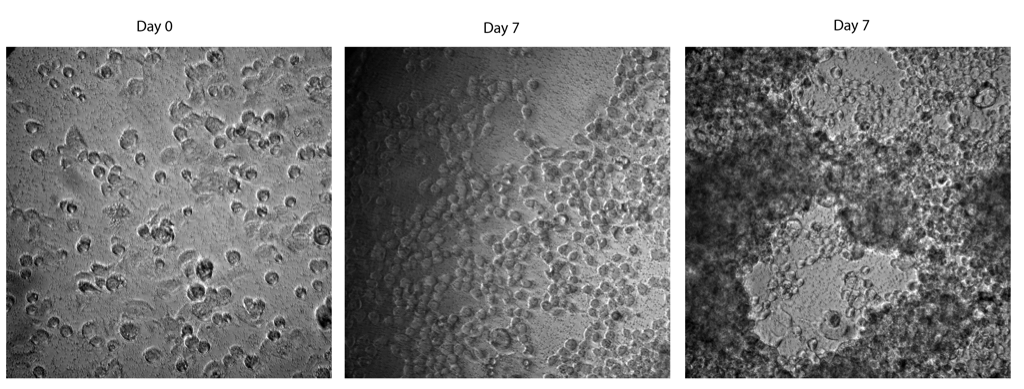
At day 0, there are mainly spherical cells. At day 7, the cells are mostly in large multi-layer clumps (right image) – the cells are out of focus because they are growing in multiple layers. The middle image is an area of lower density cells toward the edge of the PET transwell.
The same area of interest in Live/Dead fluorescence:

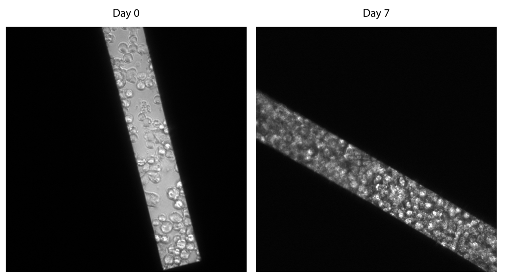
pnc-Si:
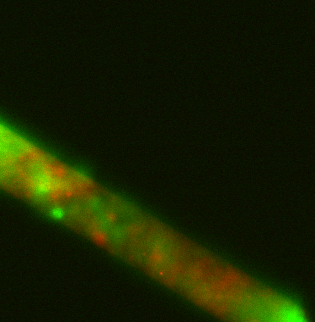
The PET images doesn’t look very good because of the large cell clumps. I notice that these cells tend to grow in multiple layers in T-75 and T-25 flasks, also, so these clumps are not necessarily a bad thing. The image of cells on pnc-Si looks terrible. I think the cells are dividing and stacking on top of each other in the well, thus creating a several cells-thick layer. It looks like there are quite a few dead cells as well, which is likely due to limited nutrient access in the middle of this multilayer. Since I seeded at a low enough density to minimize cell aggregates at Day 0 – see above image – I’m not sure how to prevent this multilayer formation.
The clumpy nature of this culture will be of no consequence if TEER values are really low. If the cells only create leaky layers on their own, but can nonetheless potentate the resistance of the endothelial cells, then we have something.