ADSC differentiation and networking in various environments
Scope:
The purpose of this experiment was to determine which conditions are best for differentiating adipose cells into endothelial cells and formation of networks. ADSCs were used for this experiment, variables included media and matrices. Geltrex and collagen were used as matrices for the cells, the bottom of the plate was used as the control. Four types of media were used: ADSC undifferentiating media, ADSC undiff media+ VEGF M200+LVES+PenStrep, and M200+LVES+VEGF+PenStrep. The ADSC media+ VEGF trial took place one passage later than the other trials.
Results:
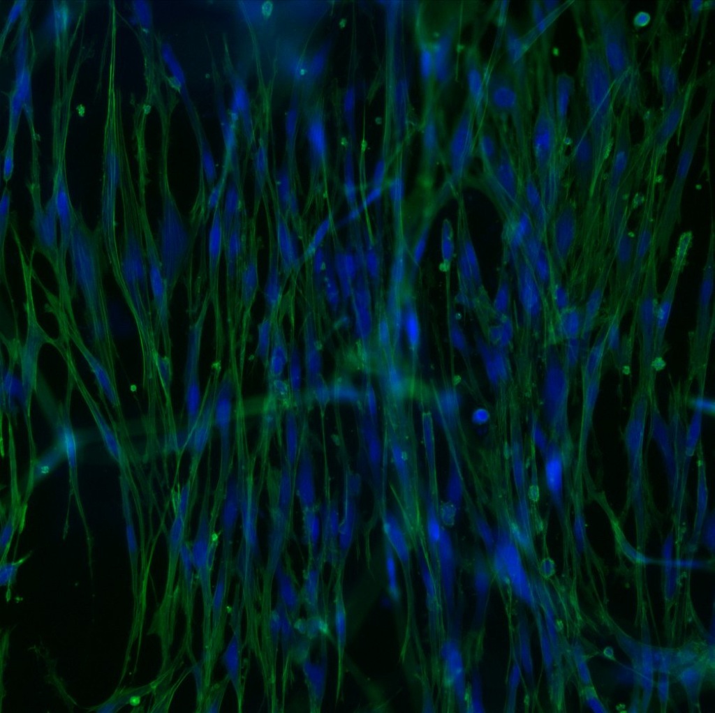
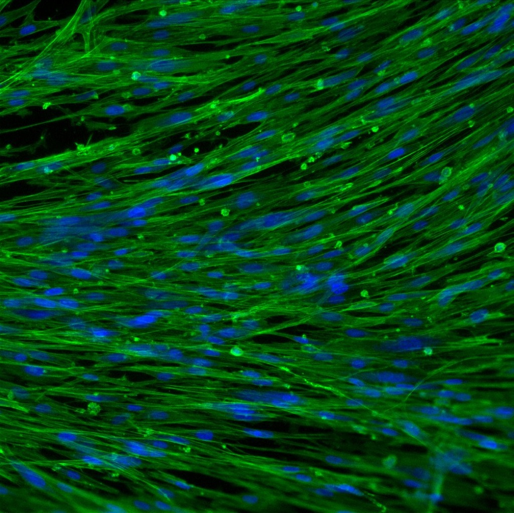
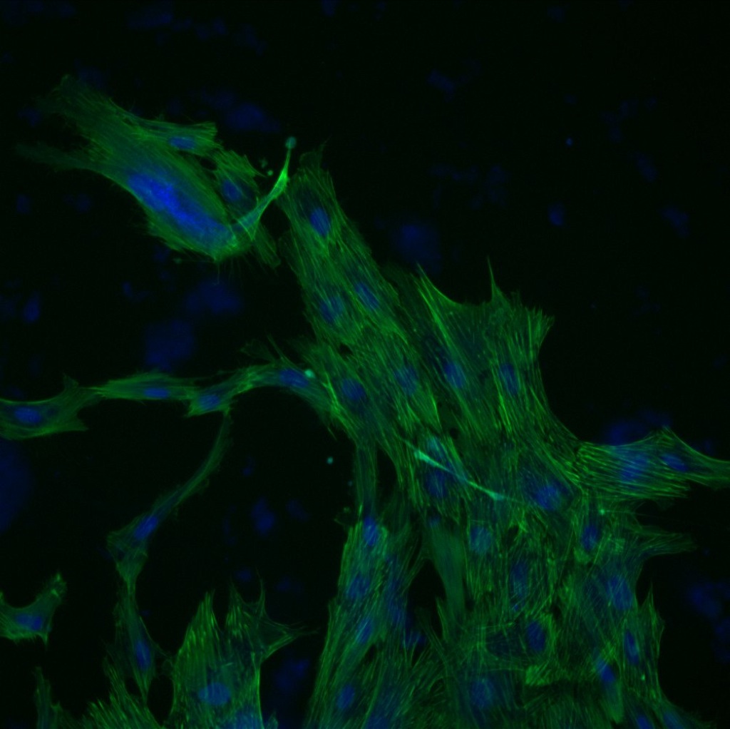
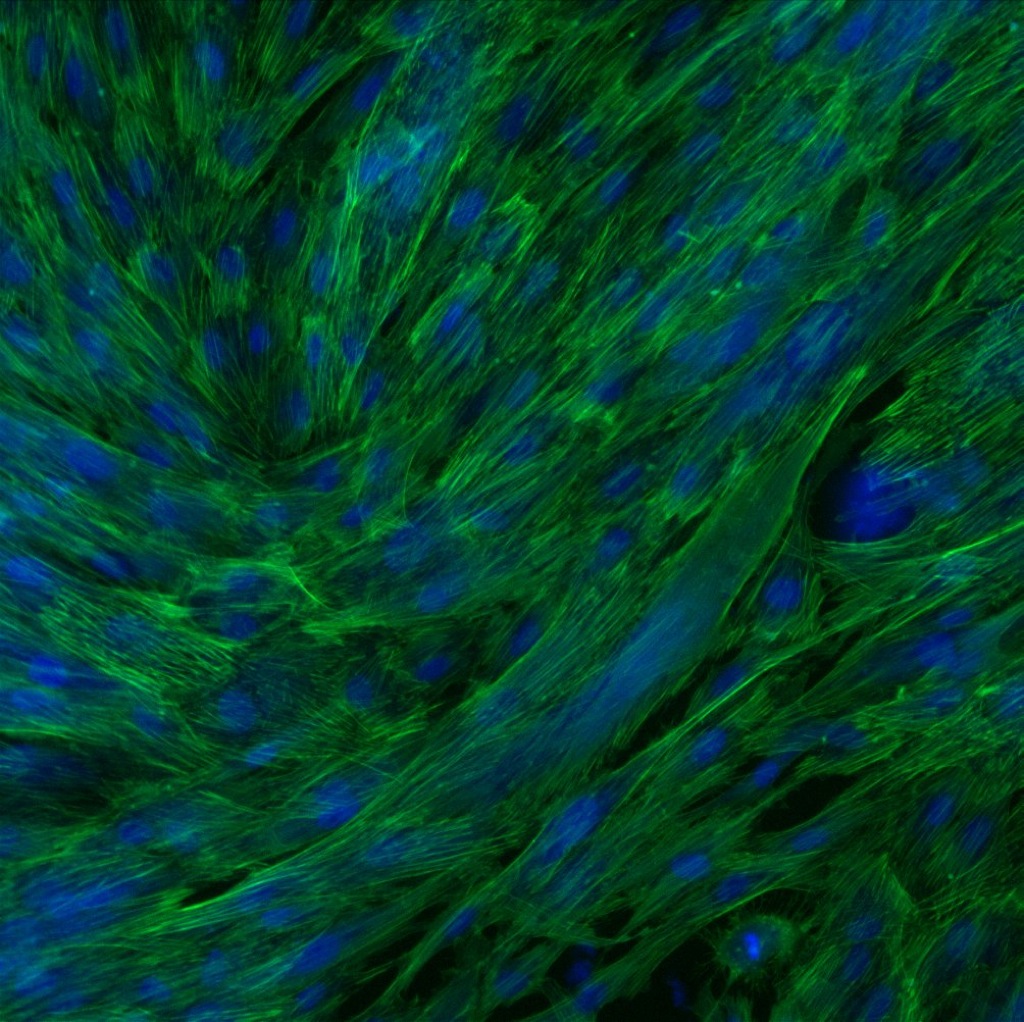
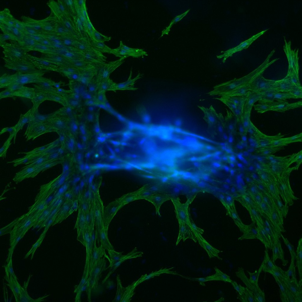
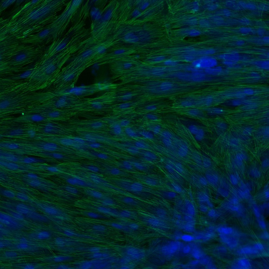
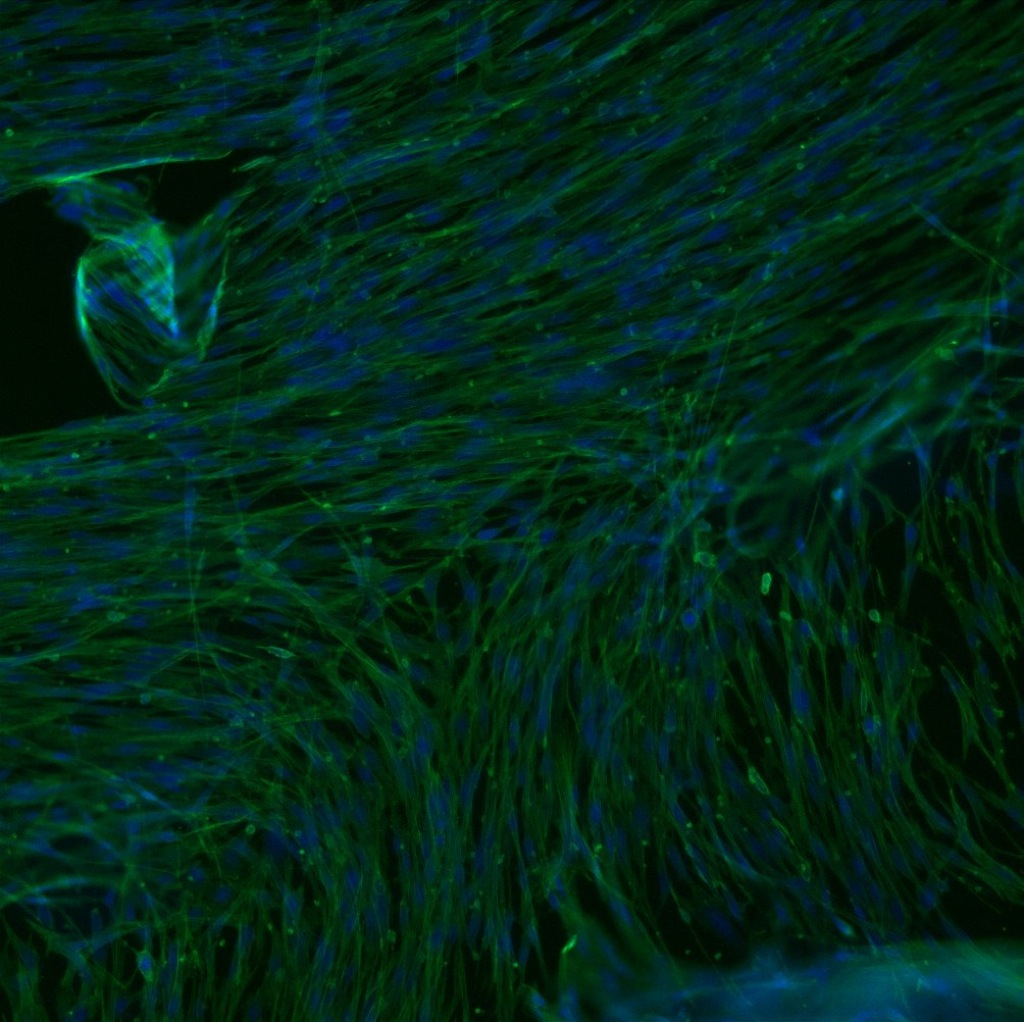
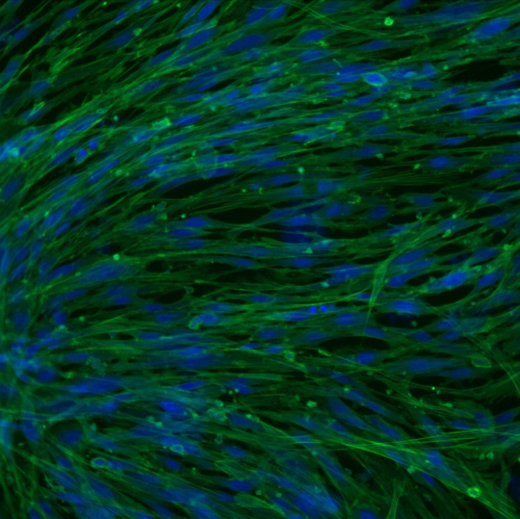
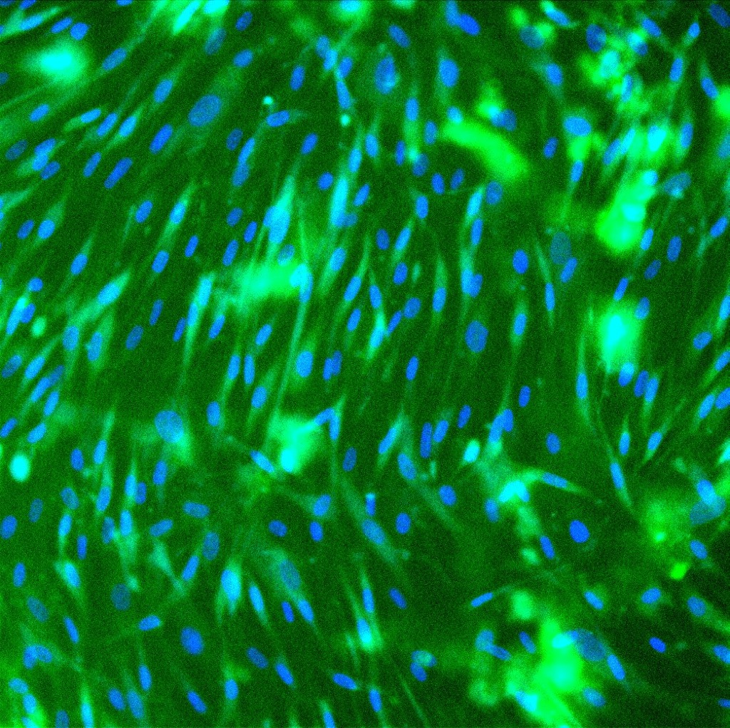
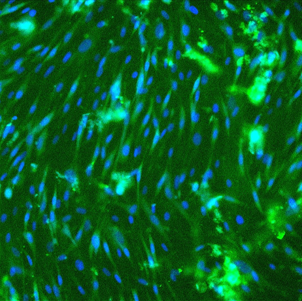
Phase images of each of the conditions were taken every 8-12 hours for 5 days. At the end of the five days cells were stained with phallodin and DAPI for actin/nucleus fluorescence or CD31 antibodies and DAPI for nucleus and endothelial cell presence. The images appeared as shown below.
Wells with Geltrex and Phallodin/DAPI stain:
| ADSC media (10x) | ADSC media+VEGF (20x) | M200+LVES+PenStrep (20x) | M200+LVES+VEGF+PenStrep (20x) |
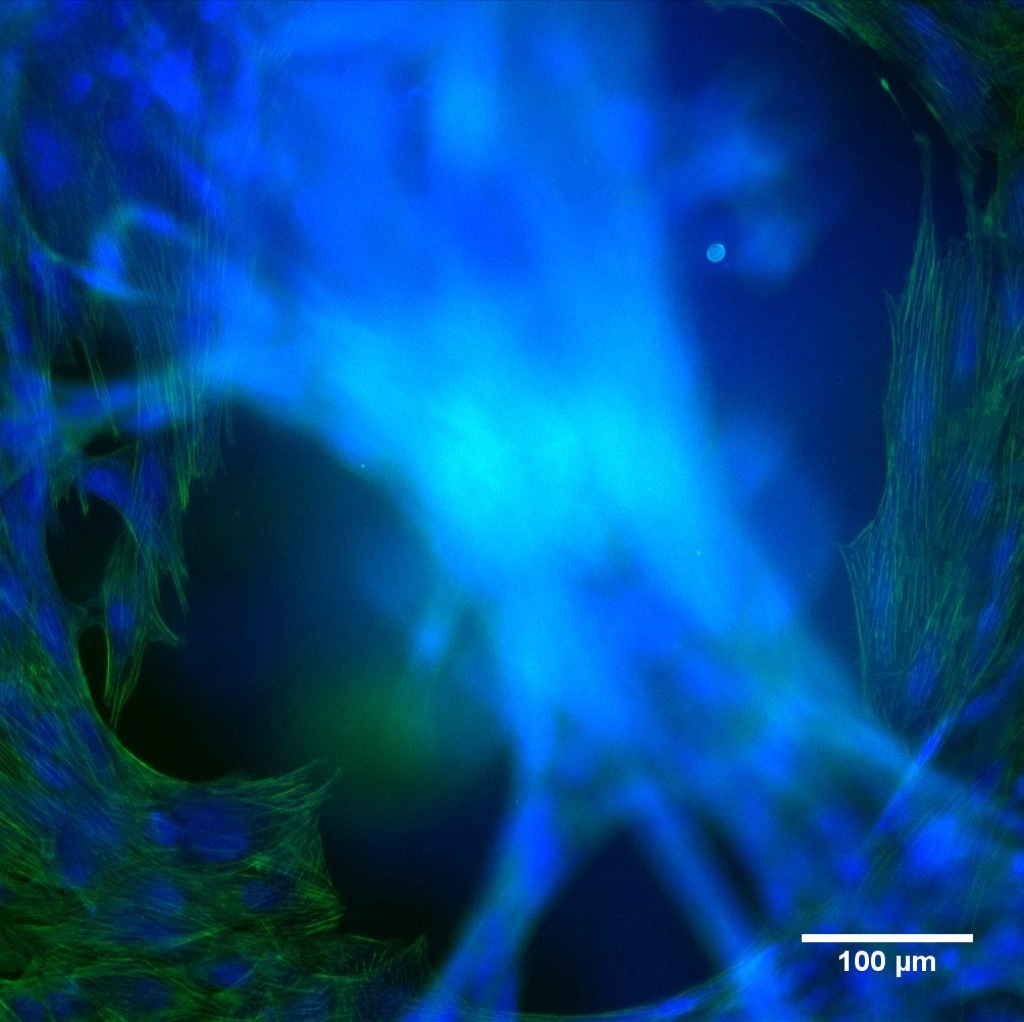 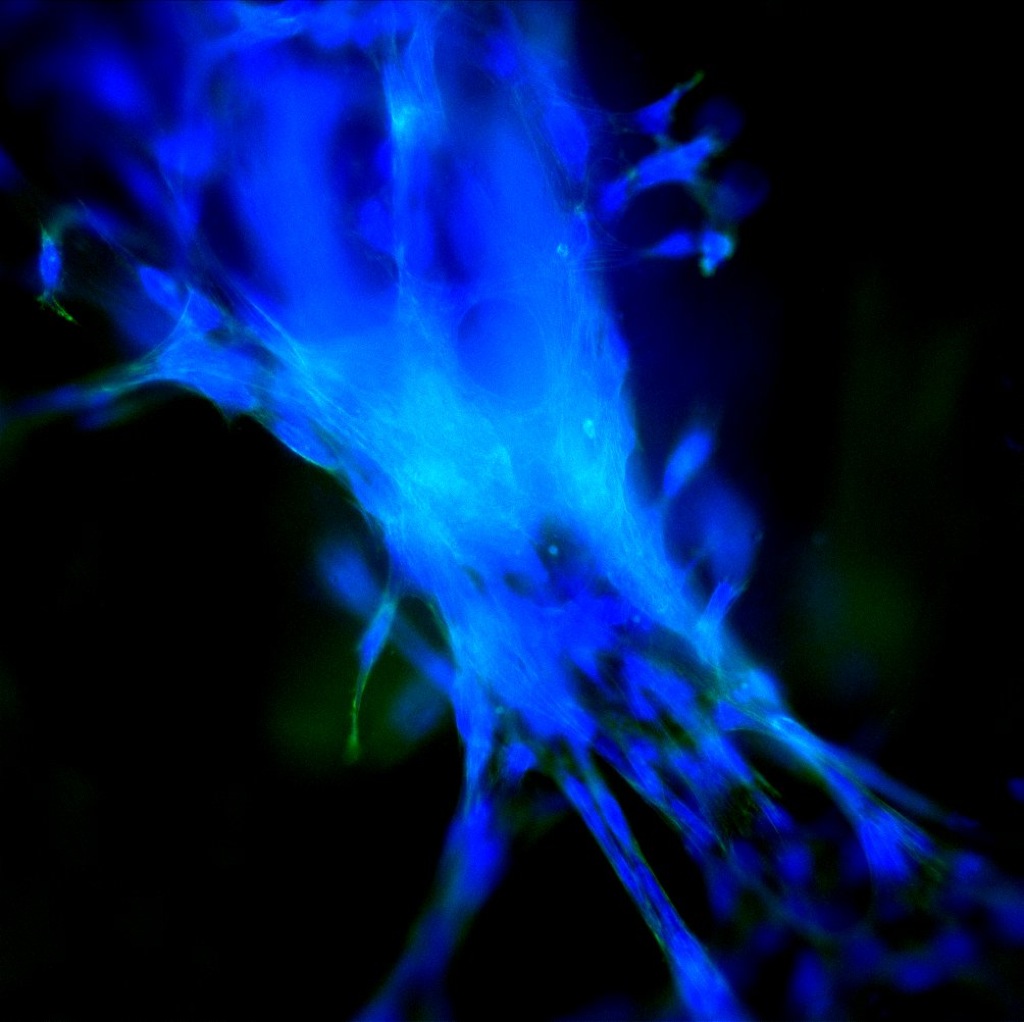
|
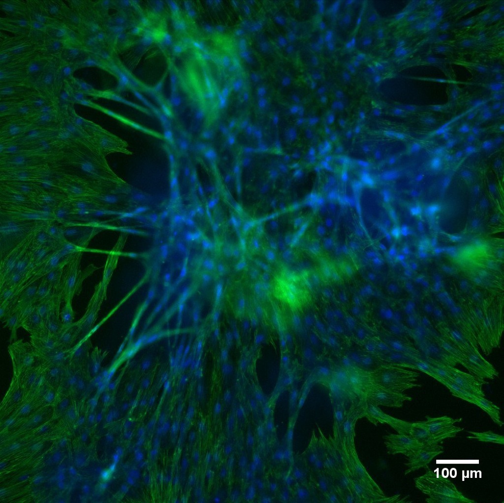 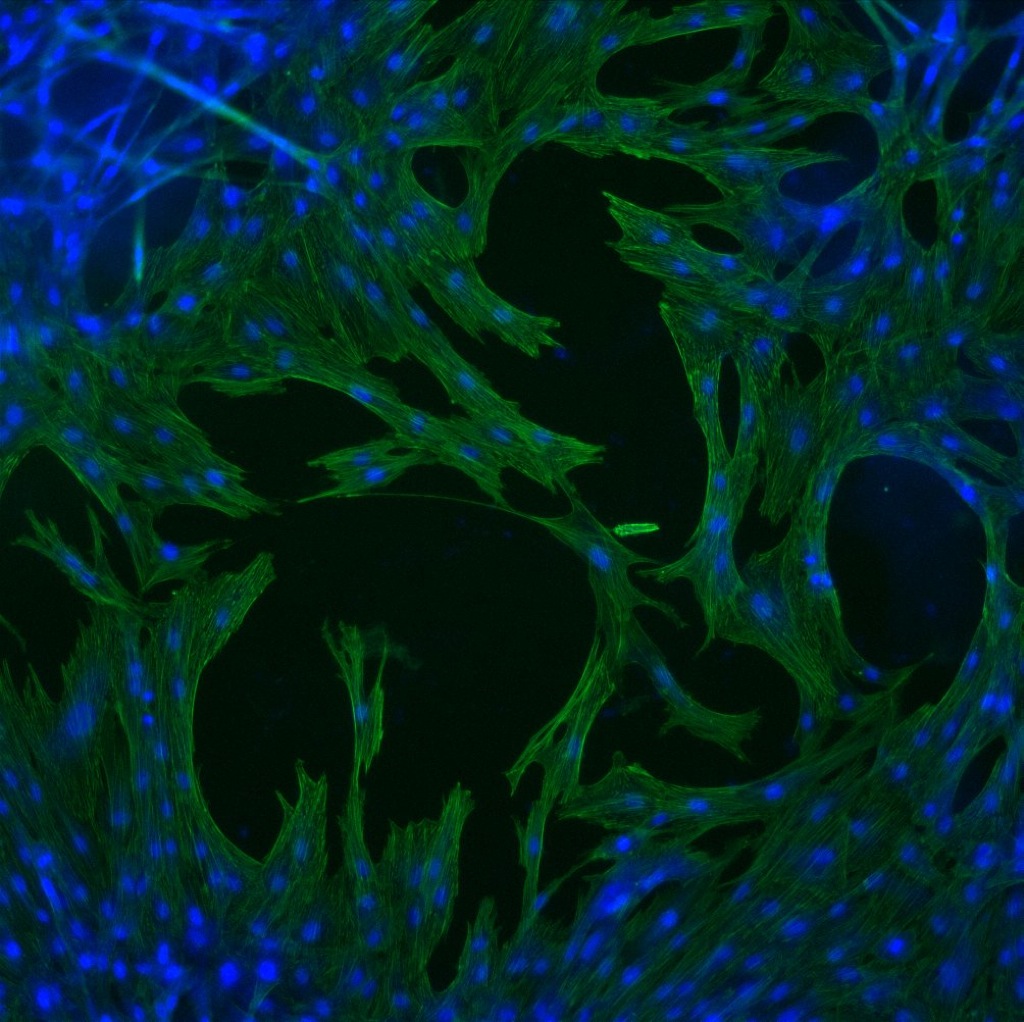
|
|
|
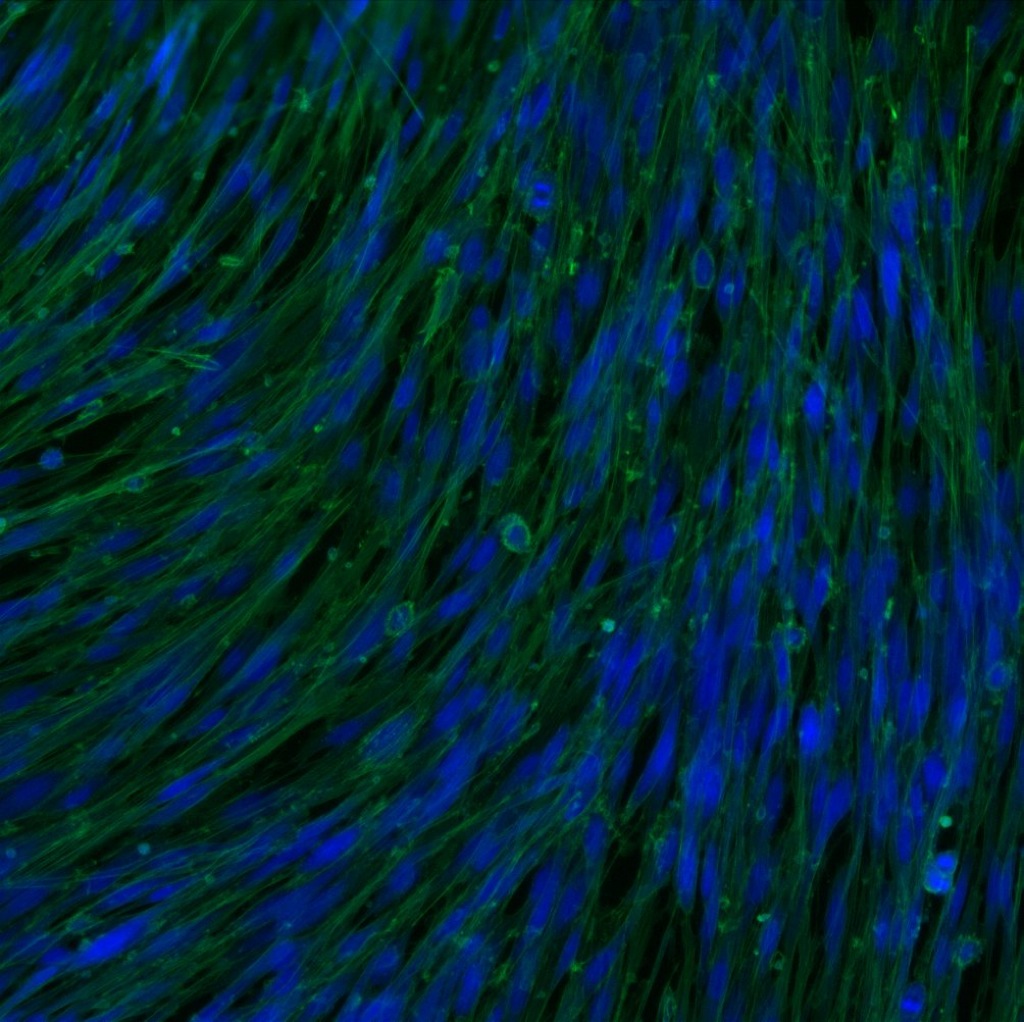
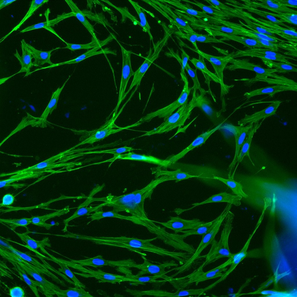
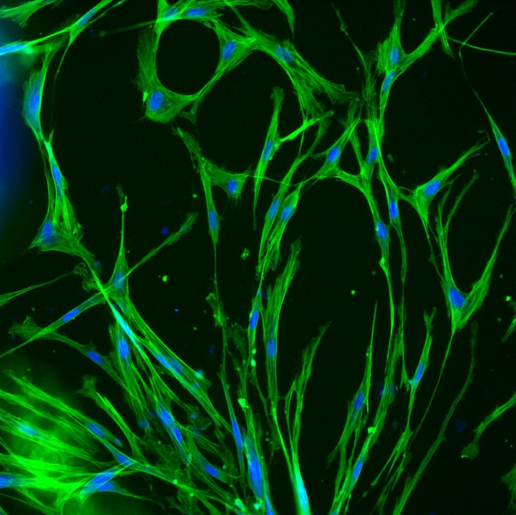
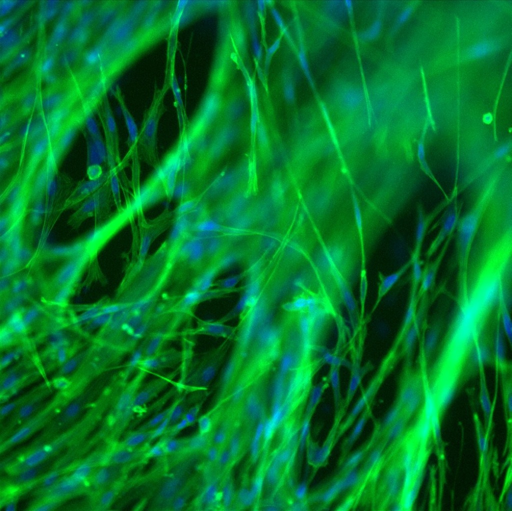
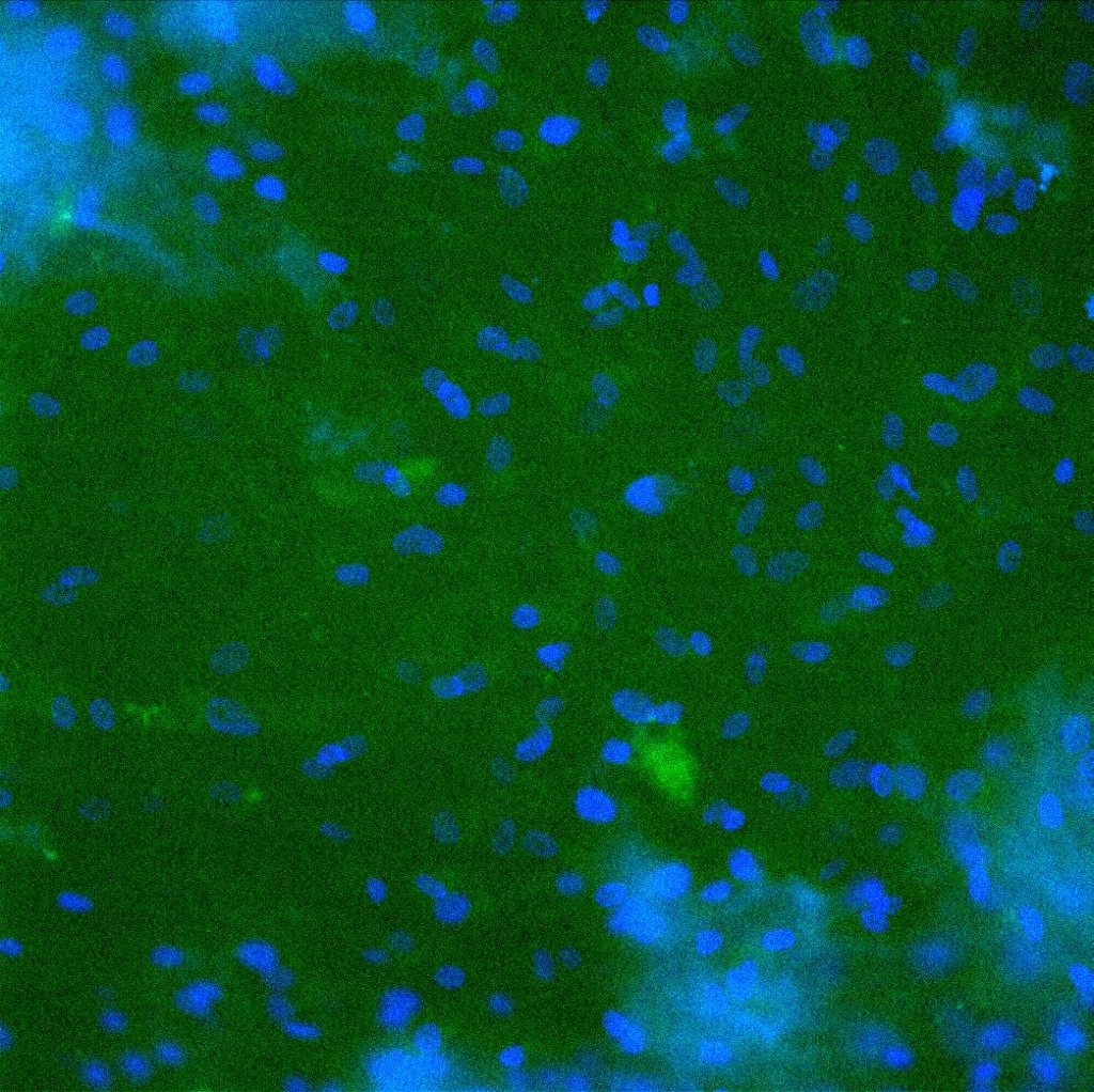
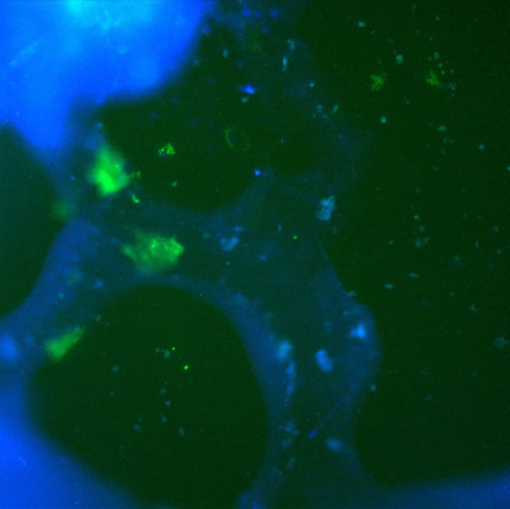
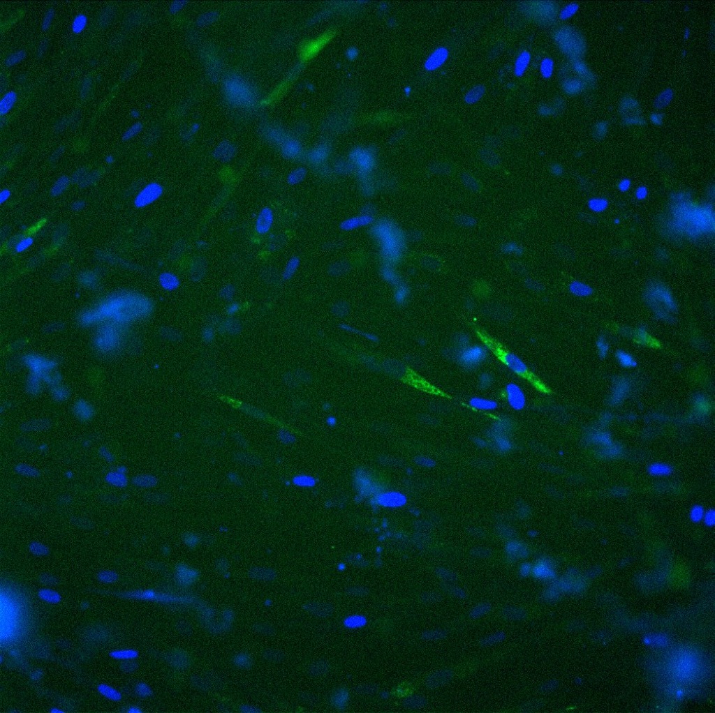
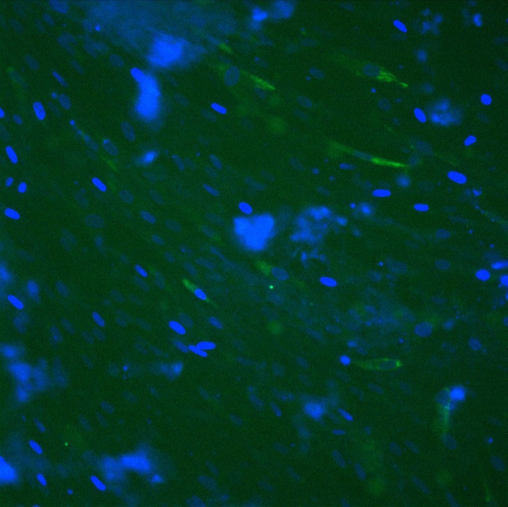
Wells with Geltrex andCD31/DAPI stain:
| ADSC media (20x) | ADSC media+VEGF (10x) | M200+LVES+PenStrep (20x) | M200+LVES+VEGF+PenStrep (20x) |
 |
 |
 |
 |
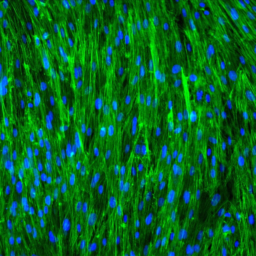
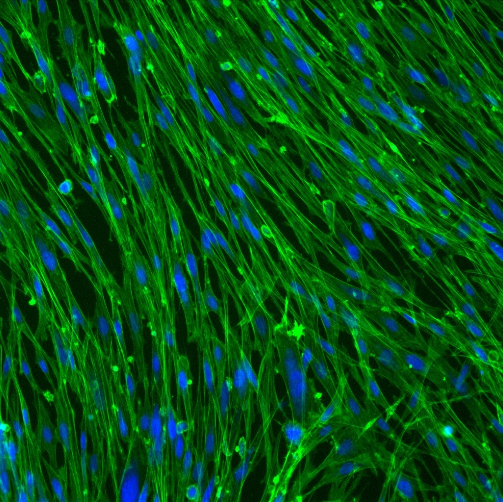
Wells with Collagen and Phallodin/DAPI stain:
| ADSC media (20x) | ADSC media+VEGF (10x) | M200+LVES+PenStrep (20x) | M200+LVES+VEGF+PenStrep (20x) |
 |
N/A | 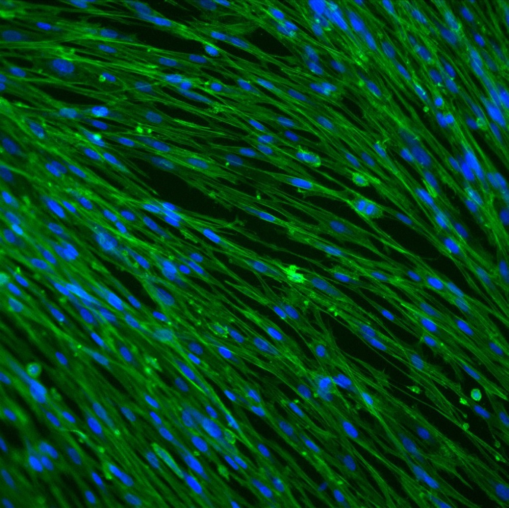 |
 |
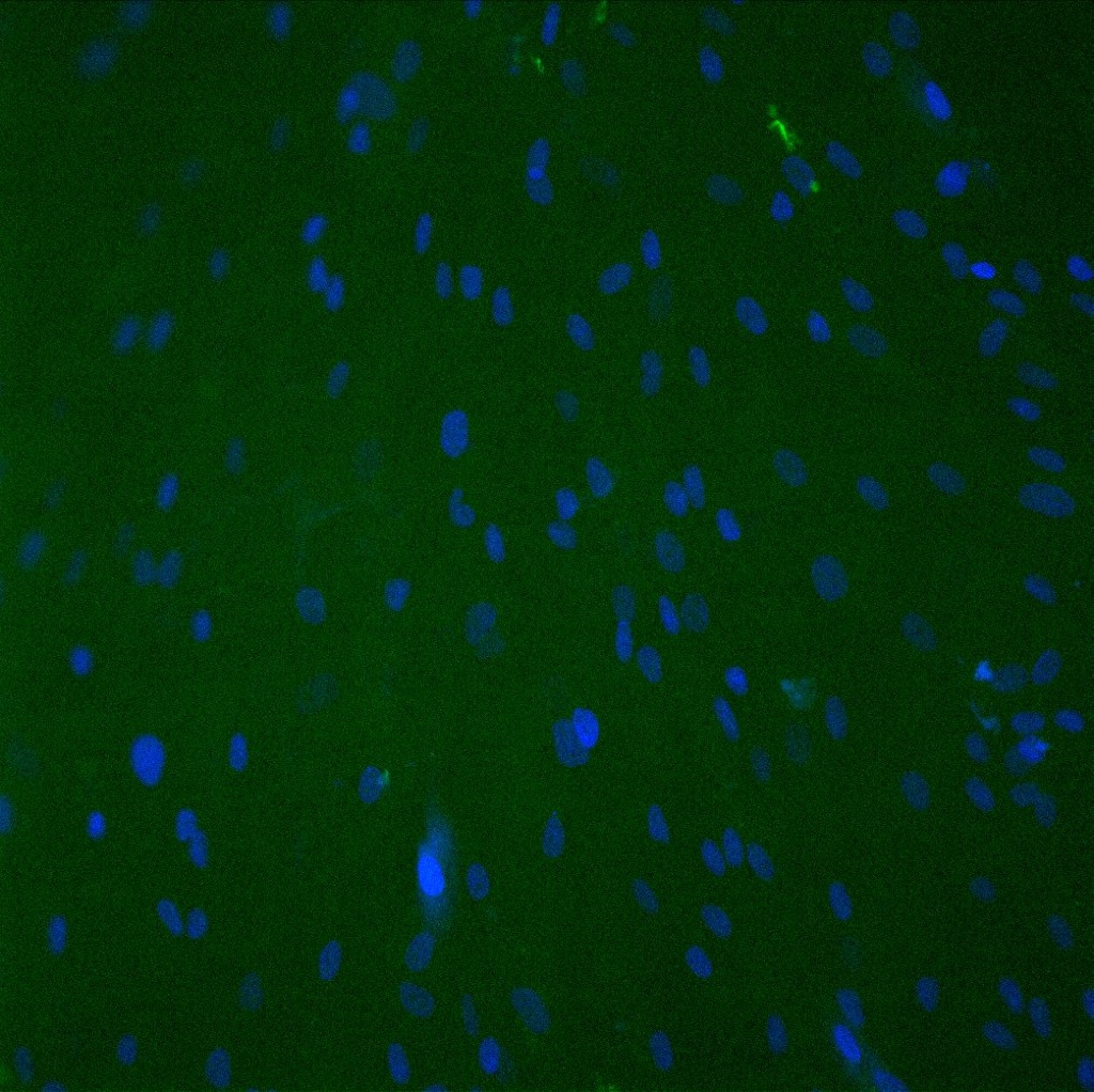
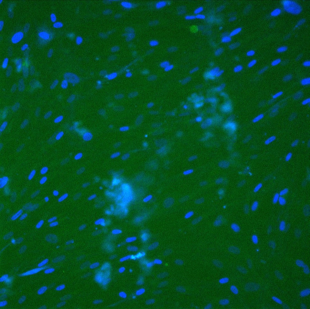
Wells with Collagen and CD31/DAPI stain:
| ADSC media (20x) | ADSC media+VEGF | M200+LVES+PenStrep (20x) | M200+LVES+VEGF+PenStrep (20x) |
 |
N/A | 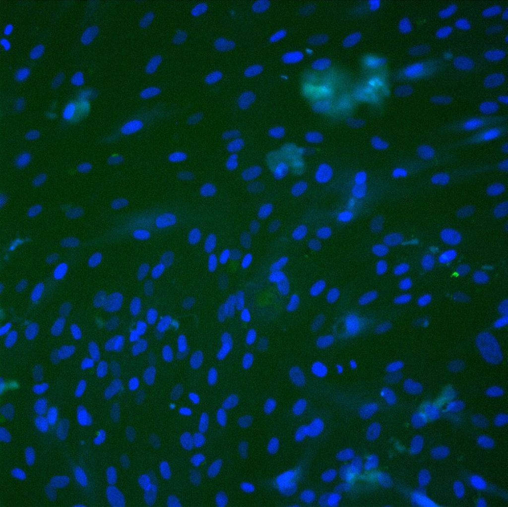 |
 |
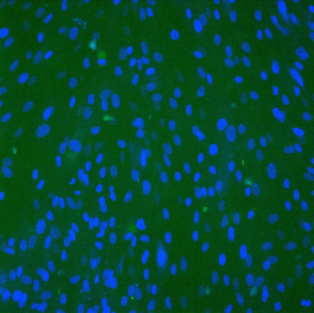
Control Wells with CD31/DAPI stain:
| ADSC media (20x) | ADSC media+VEGF | M200+LVES+PenStrep (20x) | M200+LVES+VEGF+PenStrep (20x) |
 |
N/A | 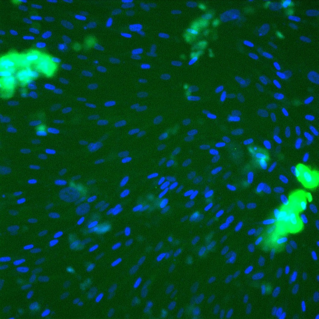 |
|
Discussion/Conclusion:
Cells in Geltrex and ADSC media appear to have more wide spread actin that interconnect with the actin of other cells. Both the HUVEC media types appear to have more narrow, stringy cells that are not as interconnected. All of the cells in Geltrex appeared to be forming networks of some sort, cells in collagen however did not appear to form any networks. Cells in a second set of wells with Geltrex and collagen were each stained with CD31 and DAPI, the DAPI stain worked and stained the nuclei, the CD31 however did not stain well and caused copious amounts of noise. This was most likely due to not washing the cells thoroughly. The stain did however appear to work well in the wells without matrices. The control well with HUVEC media and VEGF showed CD31 staining around the nuclei showing that the cells may be endothelial.
Future Experiments:
Experiments to follow include doing a more conclusive study of Geltrex and its affect on ADSCs as well as the use of VEGF with HUVEC media. The HUVEC media will be added to the cells at different time points and combined with the ADSC media to prevent shocking the cells. More care will also be taken with the CD31 stain and adequate washings will be carried out.