Blood Protein Redux
Back a long time ago, I had performed some separations with the aim of showing the usefulness of pnc-Si in hemodialysis. I attempted separations of albumin (an important protein to be kept in the blood) and cytochrome c (used in hemodialysis research to cheaply approximate the toxic protein b2microglobulin) using three different membranes. Here is the figure we came up with during this research:
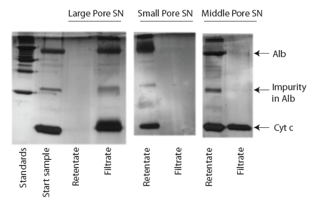
Our designation of large (266), small (277), middle (275) pore membranes were a bit off, but I think we internally accepted that small (277) and middle (275) had similar sized pores but different porosities at the time. I went back and looked at the wafer images again, and 277 and 275 are actually what I think we would refer to as having none or only background pores. All these images are film from the old TEM, but I carefully went and processed them. Here are the results:
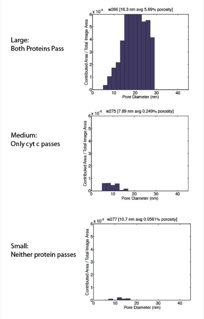
Small is not really small, just incredibly low in porosity. Passage of cyt c was repeated in a few gels with the medium membrane, so even though it may look like only background pores, there’s enough pores to let through a 3.3 nm protein. However in a couple of these gels there’s also a faint line for albumin. I can’t say we were as careful back then looking for pinholes or other problems, but there must have been a couple membranes from that wafer with pores large enough for the 5.7 nm albumin to pass (protein sizing from zetasizer).