HUVEC adhesion – pnc-Si vs. PET
I recently completed a HUVEC adhesion study with commercial PET and pnc-Si transwells. HUVEC were seeded @ 100000 cells/cm^2, allowed to attach for 5 hours (upside down format) and then stained with CMFDA. Images were acquired without autocontrast, but I did autocontrast them in ImageJ to make them brighter.
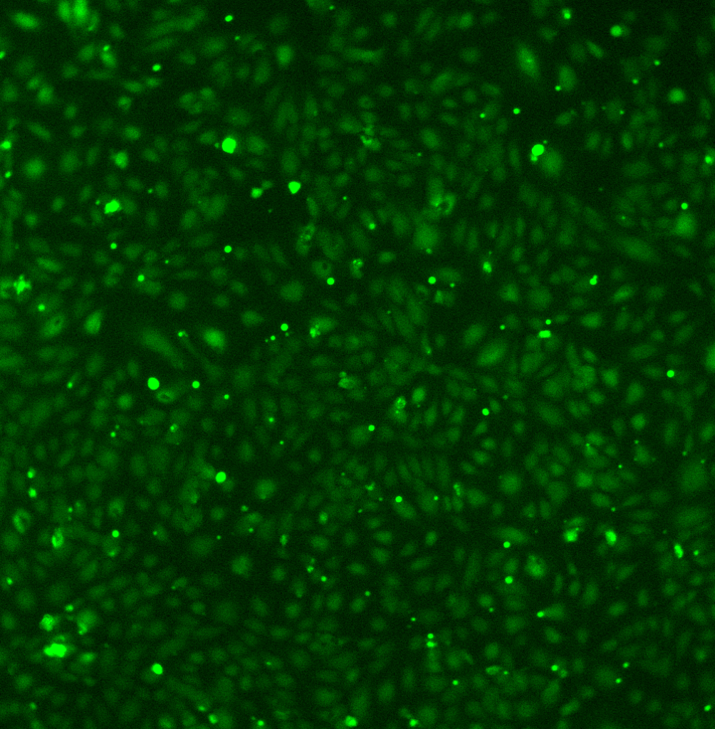
Corning Costar PET transwell:
pnc-Si transwell (with blue phase contrast overlay) – this image shows the least dense cell adhesion of the 4 I captured.
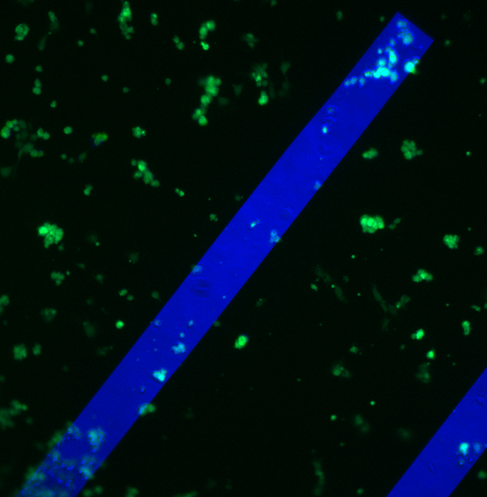
HUVEC morphology looks normal on the commercial transwells, but the cells are rounded/spindly on pnc-Si. This is the same morphology that Anant has been seeing recently (here and here). It must not be a problem with HUVEC since their morphology is good on PET transwells (and their morphology recovers after a couple days in culture). I noticed that the cell density on commercial transwells decreased considerably the closer to the membrane edge I image.
The percent adhesion values for these images (calculated from MATLAB):
Corning = 43+/- 5%
pnc-Si transwell = 27 +/- 9%
The pnc-Si value is much lower than recent posts (here, here, here). It’s possible that the very high seeding density permitted “normal” cell adhesion but then prevented cells still in suspension from adhering to the surfaces. Regardless, cell adhesion on pnc-Si is lower than that on commercial polyester transwells.
Remember that in the 2-D format we found no differences between adhesion. The spreading rate seemed normal too. So this is a problem that comes when we move to the 3-D format.
We should probably try this with 3T3s to see if we are dealing with a HUVEC – related issue.
True that adhesion and spreading were similar between pnc-Si and other substrates in 2D, but there was never a comparison to PET membranes in 2D. I agree that it’s worth doing with 3T3.