Staph Aureus Immobilized in Microchannels
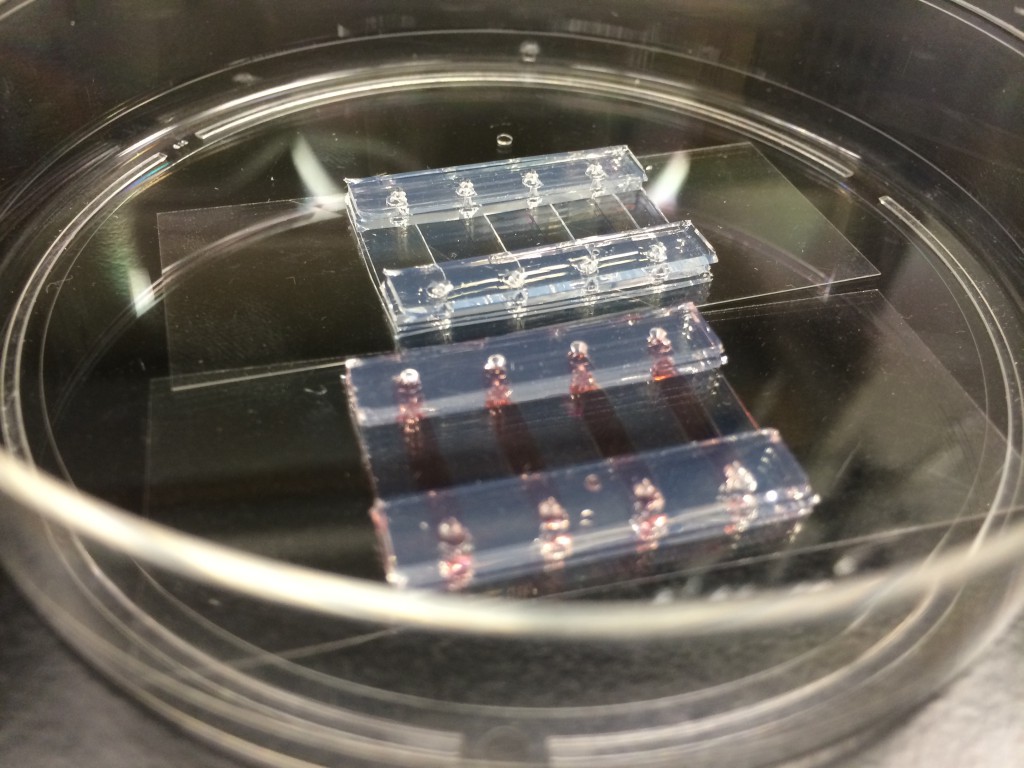
I have been developing a microsystem for Paul Dunman’s group that will allow them to flow in bacteria, immobilize them on antibodies, and then subject them to varying concentrations of antibiotics. The full system will incorporate a fluidic mixer and a NPN membrane as a way to introduce the antibiotic
I have been working with Catlyn Blanchard on this particular run of channels, and she has been great at getting the samples prepped and imaged. The microchannels (0.3 mm high by 1 mm wide) were fabricated out of silicone using the silhouette cutter, autoclaved, and sealed without membranes. Protein G (500 ug/mL) and anti-Staph antibodies (500 ug/mL) were coated in the 4 microchannels to provide attachment. Staph cells suspended in 0.8 M NaCl were stained with a GFP marker for 10 minutes (attaches to cell membrane), then introduced to the microchannels (20 uL seeded, 10^8 – 10^9 cells/mL, manually from a micropipette) and incubated for 30 minutes. Images were taken using a fluorescence scope at 10x.

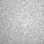
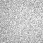
After imaging, each channel was subjected to different flow rates (20 uL/min and 100 uL/min), as a washing step. Glass capillaries were inserted in and a syringe pump was used to keep the flow rate constant. Based on the channel geometries, the shear stresses at the widest points from these flow rates were 0.2 dynes/cm^2 and 1.0 dynes/cm^2. The same fluid volume was pushed through the channels, 60 uL, to attempt to homogenize the amount of washing. The 100 uL/min channel was primed at 20 uL/min for 5 seconds, before the 100 uL/min rat was applied

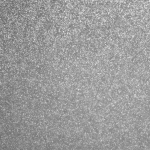
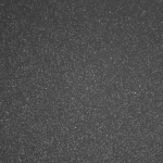
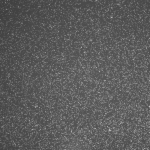
The density of cells immobilized on each channel appears to be about the same for both channels that received flow. To quantify the density, I used imageJ to threshold the image and do some simple particle counting.
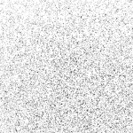

So there appear to be some differences between the two, but my opinion is that they are not very remarkable. There are less cells in the channel that was washed more vigorously, and they seem to be a little larger compared to the slower channel. Summarily, we can flow in the bugs and have them immobilized on the slides at realistic flow rates. Next, we will have to ensure that they are alive after washing, using a live/dead stain.
What is a GFP marker and how does it selectively label the cells?
Great Work!