Progress in Modelling Sepsis: Diminishing PMN chemotaxis towards fMLP gradients in µSiM Devices
First, a quick publication update. Recently we’ve published a paper on our efforts towards computationally analyzing polymorphonuclear leukocyte (PMN) trafficking on a vascular endothelium via hand-built µSiM flow cell devices. We record videos in phase contrast with a 40x objective and are able to delineate PMN state, speed, and persistence by leveraging ML algorithms and scripting. You can read this by accessing the following link:
I encourage you all to read this paper to get an idea of the abilities and limitations of our computationally assisted assay. All of the following data uses this script for analysis. Note, we take 30 minute videos (1 frame/4 seconds) and use the last 15 minutes of transmigration data as our average datapoint.
Now, onto a major update.
We have been modelling sepsis by incorporating a ‘cytomix’ cocktail (equimolar TNF-a, IL-1B, and IFN-g) into the apical or basal compartments of our µSiM flow cell devices to see how PMNs subsequently respond (Fig 1). The apical stimulation mimics endothelial exposure to circulating inflammatory factors while basal stimulation mimics inflammation originating from tissue-derived sources. As endothelial cells (ECs) exhibit apicobasal polarity, we anticipated differential PMN transmigratory responses to ECs stimulated from one side versus another depending on the level of inflammation. There’s an update on how escalating apical-sided inflammation effects PMN response, and we’ll save that for another post.
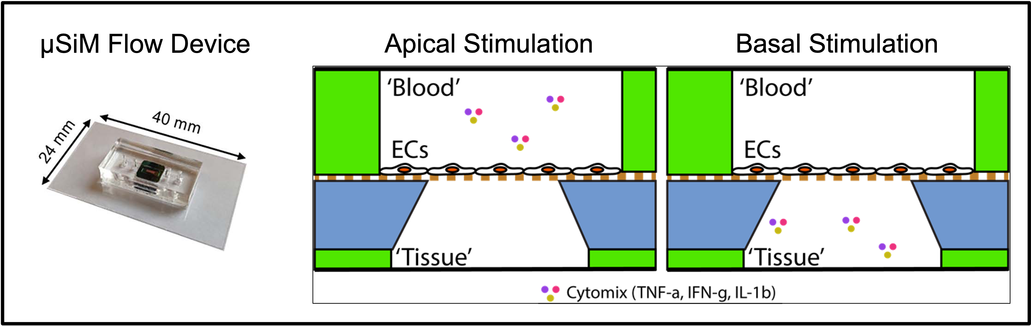
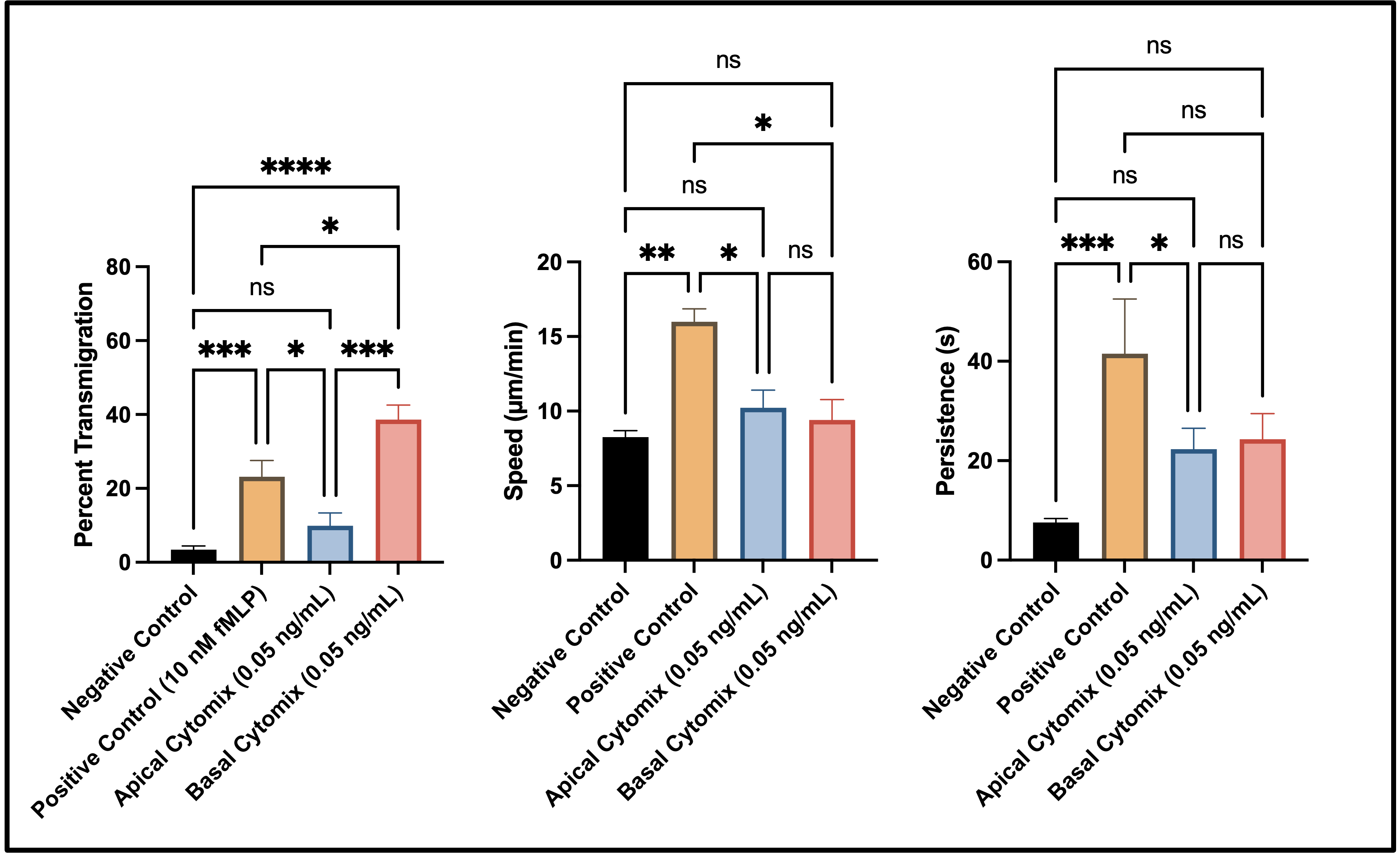
These studies were performed on two different types of ECs: Human Umbilical Vein Endothelial Cells (HUVECs) and Extended Endothelial Cell Culture Method – Brain Microvasculature Endothelial Cells (EECM-BMECs). This post will discuss HUVECs only. For HUVEC devices (seeded at 40,000 cells/cm2), we stimulated apically (n=6) or basally (n=3) with 50 pg/mL of ‘cytomix’ for 20 hours before introducing PMNs into the top channel at a concentration of 2500 cells/µL (roughly 5500 cells total for the top channel volume of 2.21 µL). For controls, one group (n=6) of devices experienced no stimulation in either channel (negative control) while another (n=6) incorporated 10 nM of N-Formylmethionyl-leucyl-phenylalanine (fMLP) into the basal compartment to act as a potent PMN chemoattractant (positive control). We expected the tissue sided stimulation to solicit the most robust PMN transmigration response, which is exactly what occurred (Fig 2). Interestingly, fMLP solicited the highest PMN speeds and persistence. Speeds for negative control PMNs and both sided stimulation groups were similar. Negative control devices displayed the lowest PMN persistence while the sided cytomix studies were similar when compared against one another.
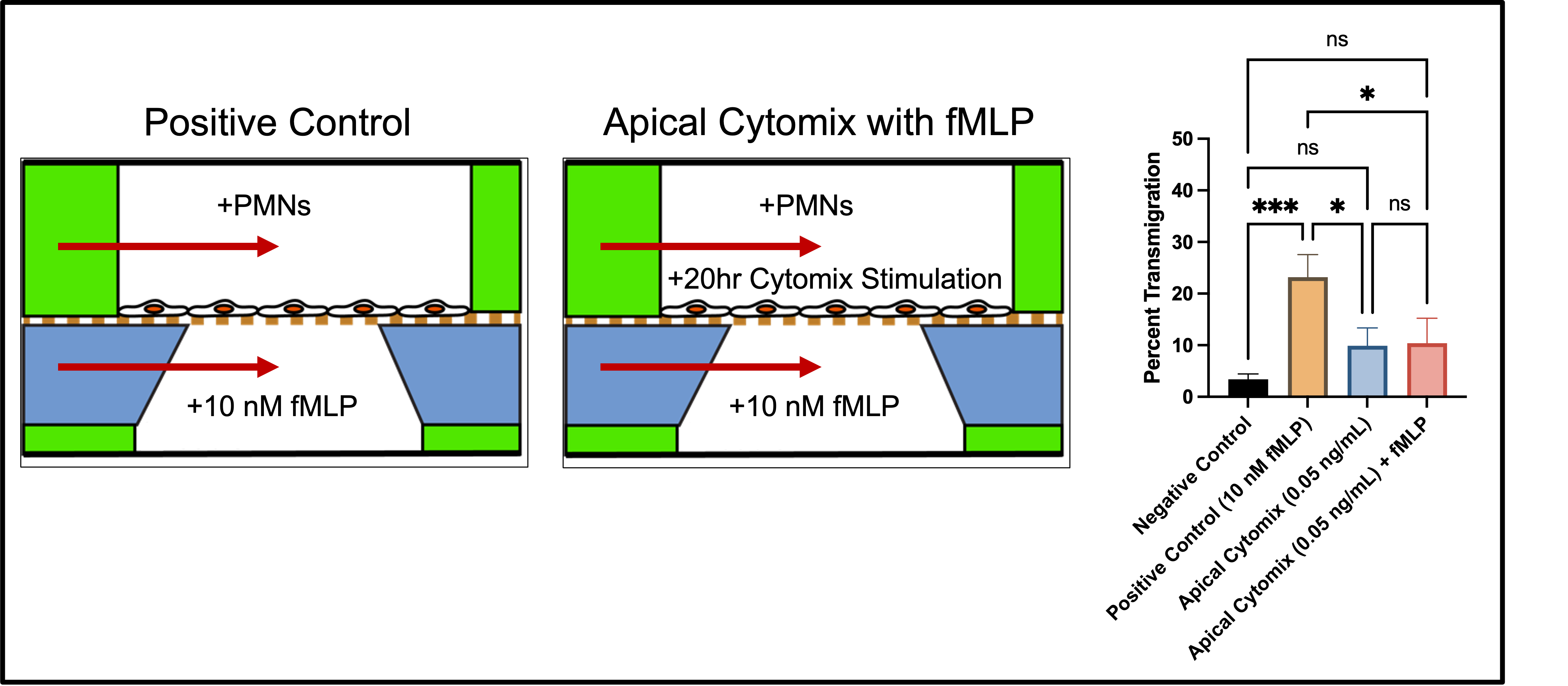
One result that stood out was the apical stimulation, which resulted in low PMN transmigratory response that was statistically similar to negative control studies. In our model, it seems as though luminal exposure to 50 pg/mL of cyotmix is not enough to overcome endothelial resilience towards PMN trafficking, however we have seen that escalating inflammation (250 pg/mL) will defeat EC apicobasal polarity in PMN transmigratory response (data not shown). In sepsis, PMNs are known to become desensitized towards fMLP due to the overwhelming presence of a variety of stimulatory factors (eNOS, IL-8, etc.). We sought to see if we could recreate this phenomena in our model (Fig 3). Interestingly, PMNs seeded into devices with EC’s (treated with cytomix, 20 hours, 50 pg/mL) failed to recover transmigratory response when fMLP was incorporated into the bottom channel. This warrants further study, and updates will be posted as additional experiments are performed.