ADSC Mechanotransduction: YAP/TAZ, adhesion complexes, and the cytoskeleton
YAP/TAZ
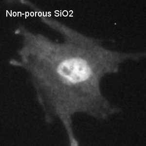
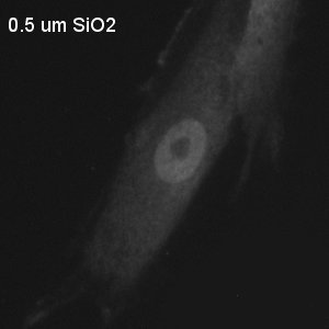
YAP/TAZ intracellular localization, and subsequent transcriptional activity, has indicated its role in pathways of mechanotransduction. Investigation of the effects of substrate stiffness on MSC growth and differentiation point to YAP/TAZ as an indicator of how cells are perceiving their mechanical microenvironment and a regulator of the cells’ response to this environment. MSCs cultured on stiff substrates exhibit nuclear localization of YAP/TAZ, while cells on soft substrates show YAP/TAZ diffusely distributed throughout the cytoplasm. Little, if any, work has been done to investigate the effects of substrate geometry, specifically contiguous stiff surface disruption, on YAP/TAZ localization.
Low-passage ADSCs were cultured on 1% Geltrex-coated SiO2 membranes for either 6 or 24 hours. ADSCs were fixed, permeablized, and stained with anti-human YAP antibody for visualization of YAP/TAZ localization.
Analysis protocol was standardized by acquiring mean intensity values from the following regions:
|
Nucleus
|
Cytoplasm
|
Background
|
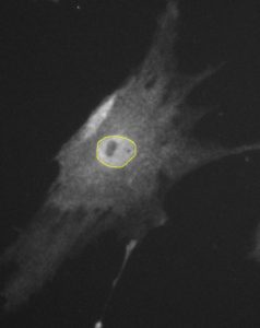
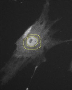
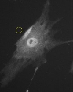
The analysis protocol was adapted from Brian Cosgrove. It is important to note that the cytoplasmic intensity was measured only within the perinuclear region, no farther than 10 µm away from the edge of the nucleus.
YAP/TAZ nuclear localization ratios were calculated by subtracting the background intensity value from both the nucleus and cytoplasm intensity values. These background-subtracted intensity values were then divided to obtain the ratio, so that ratio = (nucleus-bkgd)/(cytoplasm-bkgd).
|
6 Hours
|
24 Hours
|
|
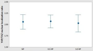
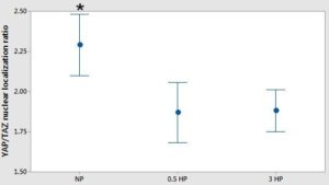
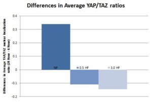
Interval were calculated using the pooled standard deviation. No statistical difference was observed at 6 hours. However, at 24 hours, the YAP/TAZ ratio in cells cultured on non-porous SiO2 was statistically different from those cultured on the 0.5 µm and 3.0 µm porous SiO2 membranes (one-way ANOVA, p<0.05). The YAP/TAZ ratio was not statistically different between the porous membranes at this time point. Differences in the YAP/TAZ ratios over time, calculated as ratio at 24 hours minus ratio at 6 hours, shows an increase in the YAP/TAZ ratio on non-porous SiO2 and a decrease on the 0.5 µm and 3.0 µm porous SiO2 membranes (student t-test indicates no significance).
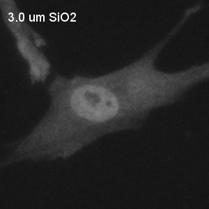
Fluorescent images of YAP/TAZ stain after 24 hours of culture:
 |
 |
 |
Adhesion complexes
Mechanisms of YAP/TAZ translocation of substrates of varying stiffness point toward integrin-mediated adhesion complexes as mechanotransducers. Specifically, differences in focal adhesion (length 2-5 µm) and fibrillar adhesion (length > 2µm) metrics have been observed between stiff and soft substrates.
Low-passage ADSCs were cultured on 1% Geltrex-coated SiO2 membranes for either 6 or 24 hours. ADSCs were fixed, permeablized, and stained with DAPI, phalloidin, and anti-human vinculin antibody to visualize the nucleus, actin cytoskeleton, and adhesion complexes, respectively. A custom MATLAB code (many thanks to Henry) was utilized to identify and quantify adhesion complexes in the 40x fluorescent images. It is important to note that some manual correction was necessary to break up aggregated adhesion complexes that the code had defined as a single complex.
 |
 |
 |
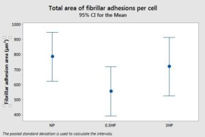 |
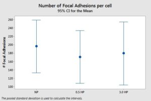
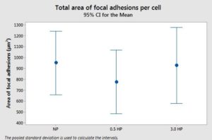
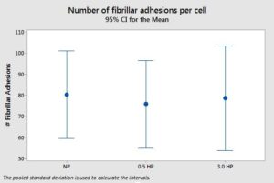
Adhesion complexes were binned by length. Focal adhesions are defined as adhesions of length 2-5 µm and are more oval in shape. Fibrillar adhesions are defined as adhesions of length > 5 µm and more resemble a straight line. No significant differences were observed in the number of focal adhesions per cell or the total area of focal adhesions per cell across the substrates (one-way ANOVA). Similarly, no significant differences were observed in the number of fibrillar adhesions per cell or the total area of focal adhesions per cell across the substrates (one-way ANOVA). Large variation was observed in the cell populations across the samples of a specific substrate and within each sample.
We hypothesize that while there is no significant difference in the size metrics (static), there may be a difference in the turnover rate (dynamic) of these adhesion complexes that contributes to how the ADSC sense the disrupted surface. Live timelapse imaging of fluorescently labeled ADSCs would be required to quantify the dynamics of complex turnover. Further investigation into the literature is required to flesh out this hypothesis.
Cytoskeleton
The organization of and tension generated by the cytoskeleton are thought to also be involved in mechanotransduction. Cells cultured on stiff substrates have distinct, organized actin stress fibers and spread easily on the substrate surface. Cells cultured on soft substrates have less distinct, slightly disorganized stress fibers. These cells do not easily spread on the substrate surface and often have a reduced cell area.
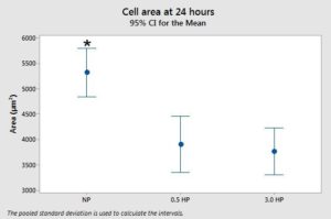
Cell spread area was calculated from the YAP/TAZ fluorescent images. ADSCs cultured on non-porous SiO2 exhibit a statistically different cell spread area than cells cultured on 0.5 µm and 3.0 µm porous SiO2 membranes.
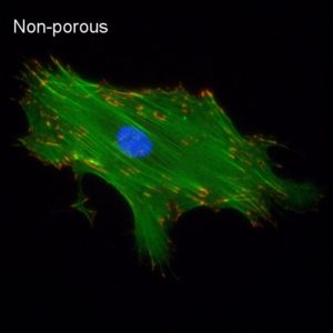
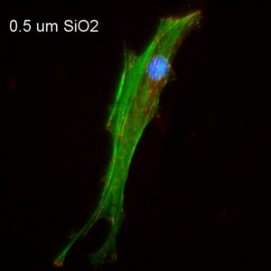
Differences in cell morphology can be observed between the non-porous and porous membranes.
 |
 |
Cytochalasin D is a compound that disrupts actin filaments and inhibits actin polymerization. Treatment with CytoD disrupts the cytoskeletal tension which would normally communicate to the cell that it is on a stiff substrate. Subsequently, the cell perceives that it is on a soft substrate and YAP localizes to the cytoplasm.
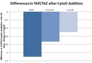
Low-passage ADSCs were cultured on 1% Geltrex-coated SiO2 membranes. The ADSC media was supplemented with 1 µM CytoD 1 hour after seeding. After 24 hours, the cells were fixed, permeabilized, and stained for anti-human YAP antibody and analyzed as previously described. Statistically significant decreases in YAP/TAZ nuclear localization were observed on all substrates (student t-test).