Fabrication update and functionalization plans
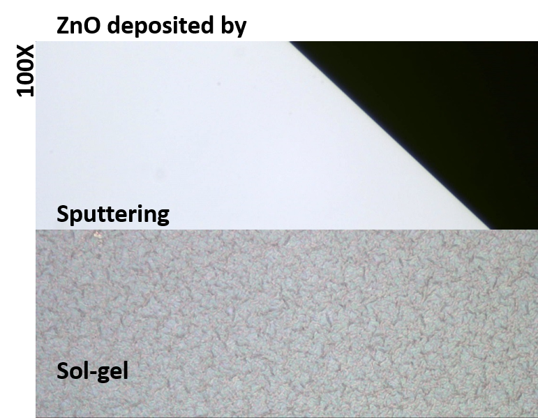
Thanks to UR cleanroom and Jim Mitchell, we finally have a better way to deposit the ZnO film that our fabrication process uses as sacrificial layer. Below is a quick comparison of the previous ZnO deposited by sol-gel and the new ZnO deposited by sputtering at UR. Hopefully, if you look close enough (or far away) you can see the difference.
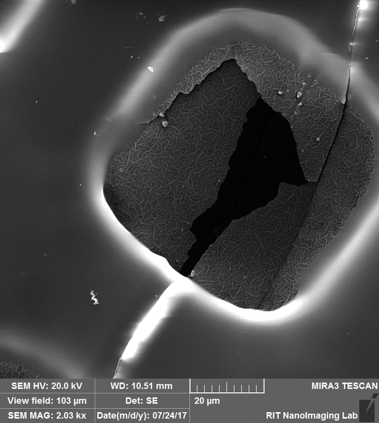
We completed the second run of the NSL fabrication process and released the membrane. In this case we were using the old ZnO, so the TEOS membrane still had those large features resulting from depositing conformally to the ZnO. There was also some overexposure of the SU-8 pattern which made it look thicker. Below you can see some SEM pictures of the released membrane that Aslan took for us.
The picture below was taken from an area where the membrane is curling, so it is essentially a tilted view. You can again observe the large features imprinted on the TEOS (from the ZnO), that causes the beads to shift and move out of place (this is an old sample, we got a lot better on bead assembly). Although not completely etched through, you can see the pores (circled).

Here is a picture taken from a broken area, you can almost see some through pores.

Below are two zoomed out views of the previous area where you can see that the tear probably originated in the SU-8 and transferred to the membrane. Also, the pictures shown here correspond to a small area close t the edge of the 2×2 inch sample.

We are currently working on the third fabrication run with the new and improved ZnO, however the PECVD at RIT is not having a great few weeks and is currently down.
Moving forward…..
The plan for Cody’s thesis is to functionalize the fabricated SiO2 membrane with a temperature responsive polymer – Poly(N-isopropyl acrylamide) or pNIPAM. Below the lower critical solution temperature (LCST), NIPAM swells, and above the LCST, NIPAM collapses. (see first schematic, as taken from Matsuzaka, N. et. al. 2013). The first goal is to release captured beads from the membrane using the LCST transition property of this polymer. The figure below illustrates the transition, and the source paper shows cell sheet detachment from a solid surface using the same polymer.