NIH 3T3 and Human Dermal Fibroblast wound healing
For the past few weeks, Henry and I have been working with human dermal fibroblast cells and 3T3 mice fibroblast cells. We have been working to quantify wound recovery in hopes to be able to incorporate exosomes from different cell lines to see the effects on wound healing. The NIH 3T3 cells are small cells that are very mobile and people often attempt to characterize their migration. In contrast, the human dermal cells are much larger and longer and tend to move slower. We purchased a dye to be able to better visualize the cells: SiR DNA. It targets the DNA and allows us to better see the cells and will allow us to create a more accurate Matlab code to characterize movement and wound recovery. Due to the different in intensity difference between the light caused by the dye the background, we obtain a greater contrast which we can then use to quantify the data.
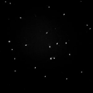
Dermal fibroblasts, migration experiment 3, geltrex coating, SiR DNA
This is a quick example of an image of human dermal fibroblasts with SiR DNA dye. The cells are easily distinguishable and they are easy to track with Matlab code.
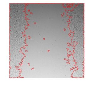
To ensure that the dye did not affect the cells in a negative way we did an experiment to compare cells with and without SiR DNA. We did the experiment for both hdFb (human dermal fibroblasts) and 3T3. We did a scratch wound assay where we imaged the cells every 15 minutes. The scratches were created with a p200 tip, a method used by many other labs. We created short movies for each well to visualize the wound healing. We were able to create a MAtlab code that quantified the wound closing from DIC images. Although the program may not be as accurate as one that uses the contrast created by SiR DNA to quantify the wound healing, we needed to create a way to determine that the dye had no effect on cell movement. The program we created uses boundary and edge detection and creates a line the show the progress of the wound healing.
Scratch wound assay, dermal fibroblast, geltrex coating, SiR DNA
Scratch wound assay, 3T3 fibroblast, geltrex coating, SiR DNA
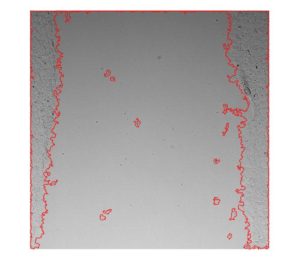
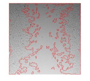
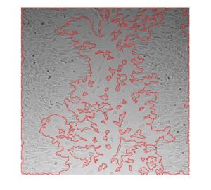
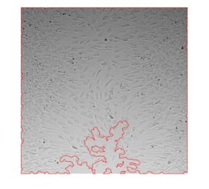
Images from the Matlab code that quantifies the wound area of 3T3 cells, SiR DNA, no coating
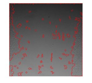
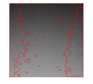
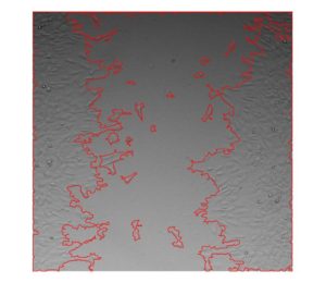
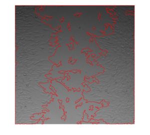
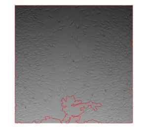
Images from the Matlab code that quantifies the wound area of 3T3 cells, no SiR DNA, no coating
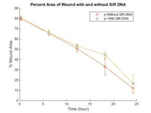
The next step was to determine whether the SiR DNA had an effect on the cells. Visually, the two scratch wound look almost identical. We created a graph and performed a 2 sample T-test and determined that the 3T3 cells with and without DNA were not significantly different.