Who’s Your Cabby? Analyzing the Roles of Haptotaxis and Durotaxis on Staphylococcus Aureus Invasion of the Osteocyte Lacuna-Canicular Network
As discussed in a previous post of mine, I am currently working on a project to develop an in vitro mimetic of the osteocyte lacuna-canicular network (OLCN) to improve the ability to predict Staphylococcus Aureus invasion and colonization of the OLCN during osteomyelitis. The purpose of this post is to provide more background behind Aim 1 of that post. Aim 1 pertains to the roles of haptotaxis and durotaxis on the invasion of the OLCN by Staphylococcus Aureus. Below I will provide definitions for both and examples of how either cell migration type could affect the ability of staphylococcus aureus to propagate into the OLCN.
Haptotaxis
Haptotaxis is a type of cell migration relating to extracellular adhesion gradients. In a physical sense, it is defined as the directional movement of cells up an adhesion gradient from a less adherent to a more adherent surface1. These adhesion gradients can be formed from adhesion sites or surface bound chemoattractants2. In a genetic sense, it is defined as motility guided by three-dimensional extracellular matrix organization3-5. Since our work concerns the physical sense, we will use the first definition.
Study Design
In our study, we want to present various surfaces which exhibit a range of adhesive properties to Staphylococcus Aureus. To accomplish this, we will use bare silicon nitride (neutral adhesion), PEG (adhesion prevention), and PLL (adhesion promotion) to create a ‘measuring stick’ with which we can compare haptotaxis of Staphylococcus Aureus on a membrane coated with hydroxyapatite. Hydroxyapatite is the main inorganic constituent of human bone, so it should provide a surface that is more physiologically relevant to bone. The ability or inability of Staphylococcus Aureus to propagate across each of these membranes will give us clues as to the role that haptotaxis plays on the propagation of Staphylococcus Aureus in the OLCN.
Durotaxis
Durotaxis is a type of cell migration relating to extracellular stiffness. Is is defined as the directional movement of cells up a stiffness gradient from a less stiff to a more stiff surface6-8.
Study Design
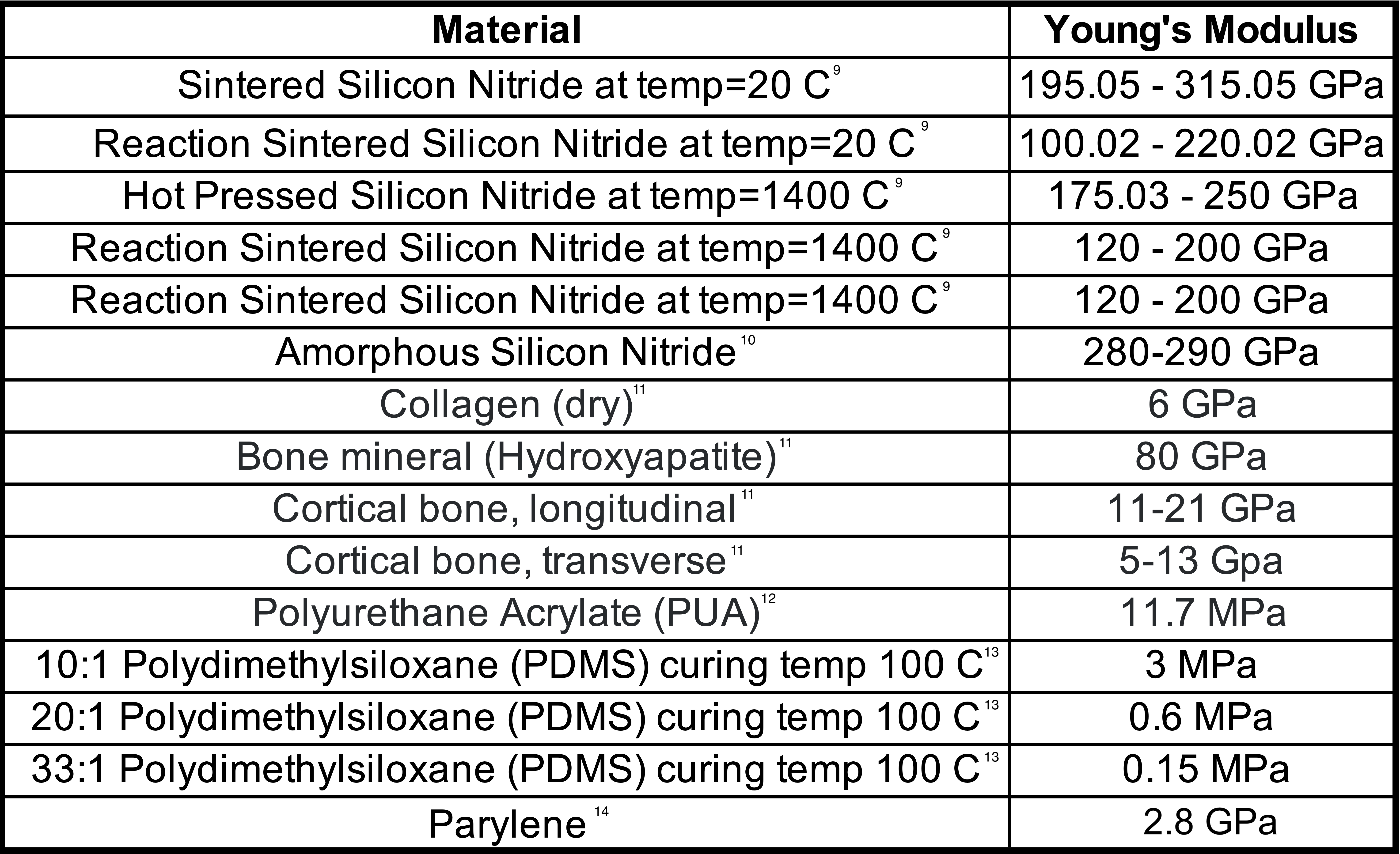
In this study, we want to present various surfaces which exhibit a range of stiffnesses to Staphylococcus Aureus. To accomplish this, we will use bare silicon nitride, hydroxyapatite coated membranes, PDMS coated membranes, and PUA coated membranes. Their Young’s Modulus are shown in the table below. By presenting a wide range of surface stiffness we will again try to tease out the role of durotaxis on the propagation of Staphylococcus Aureus in the OLCN.
References
1. Carter, S. Haptotaxis and the Mechanism of Cell Motility. Nature, 213, 256–260 (1967). https://doi.org/10.1038/213256a0
2. Wen, Jessica H et al. Haptotaxis is cell type specific and limited by substrate adhesiveness, Cellular and molecular bioengineering, 8(4), 530-542 (2015). doi:10.1007/s12195-015-0398-3
3. Masters Elysia A. et al. Staphylococcus aureus Cell Wall Biosynthesis Modulates Bone Invasion and Osteomyelitis Pathogenesis, Frontiers in Microbiology, 12, (2021). doi: 10.3389/fmicb.2021.723498
4. Hsu, S., et al. Effects of shear stress on endothelial cell haptotaxis on micropatterned surfaces, Biochem. Biophys. Res. Commun. 337, 401–409, (2005). doi: 10.1016/j.bbrc.2005.08.272
5. Oudin, M. J., et al. Tumor cell–driven extracellular matrix remodeling drives haptotaxis during metastatic progression. Cancer Discov. 6, 516–531, (2016). doi: 10.1158/2159-8290.cd-15-1183
6. Lo, Chun-Min et al. Cell Movement Is Guided by the Rigidity of the Substrate, Biophysical Journal, 79(1), 144–152 (2000). https://doi.org/10.1016/S0006-3495(00)76279-5
7. Sunyer, R. et al. Collective cell durotaxis emerges from long-range intercellular force transmission, Science, 353, 1157–1161 (2016). doi: 10.1126/science.aaf7119
8. Sunyer, R. et al. Durotaxis, Curr. Biol., 30, R383–R387 (2020).
9. MEMSnet, Material: Silicon Nitride (Si3N4), https://www.memsnet.org/material/siliconnitridesi3n4/
10. Khan, A. et al. Young’s modulus of silicon nitride used in scanning force microscope cantilevers. Journal of Applied Physics, 95(4), 1667–1672 (2004). https://doi.org/10.1063/1.1638886
11. University of Cambridge, Mechanical properties of bone, https://www.doitpoms.ac.uk/tlplib/bones/bone_mechanical.php
12. Li, Jinhai et al. Urethane-acrylate polymers in high-resolution contact printing. Journal of Materials Chemistry. C, Materials for Optical and Electronic Devices, 4(19), 4155–4165 (2016) https://doi.org/10.1039/c6tc01125j
13. Wang, Z. et al. Crosslinking effect on polydimethylsiloxane elastic modulus measured by custom-built compression instrument. Journal of Applied Polymer Science, 131(22), (2014) https://doi.org/10.1002/app.41050
14. MIT.edu, Material: parylene, http://www.mit.edu/~6.777/matprops/parylene.htm