Cancer Cell Migration and Wound Healing
A few weeks ago, Henry and I ran a migration/wound healing experiment using two cancer cell types: T24 (bladder cancer) and MDA-MB-231 (breast cancer). MDA cells are highly migratory, while T24 cells are less so. We wanted to test if the exosomes from MDA cells would cause T24 cells to become more migratory. This was accomplished by creating co-culture devices that split each well of a 24-well plate in half, seeding MDA on one side of the divide and T24 on the other. Some of these wells allowed for complete transfer of proteins and exosomes between the two cell lines. Other samples contained a membrane that blocked the transfer of exosomes between the cells but still allowed for the transfer of most proteins. Results from this experiment showed that T24 cells in samples without a membrane did better than those with a membrane. Therefore, migratory cell exosomes potentially cause less agressive cancer cell lines to become more migratory.
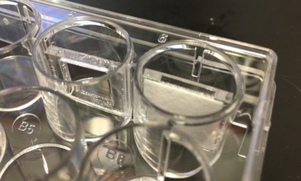
The co-culture devices were created by laser cutting acrylic into rectangular walls. Each wall piece had another rectangular hole in the middle. While some holes were left open to allow complete transfer of exosomes, others were covered with a PES dialysis membrane (fig. 1). These membranes block exosome transfer while allowing most proteins to pass through.
Cells were seeded in each well, with MDA on one side and T24 on the other. Media used for each well was 50/50% RPMI (T24 cell media) and DMEM (MDA cell media). Wounds were created in T24 cells using a pipette tip. Images of each wound area were taken every 15 minutes, along with the areas to the left and right of the wound. Cell tracking movies were created in MATLAB using these image sequences. The speed, mean squared displacement (MSD), directionality ratio, and cell density recovery were also calculated using MATLAB (fig. 2). Significant evaporation occurred between 24 and 48 hours, therefore data was only analyzed between 0 and 24 hours.
Below is a presentative movie comparing the co-culture with and without the membrane for exosome exclusion:


Figure 3. full data set including experiment using conditioned media.
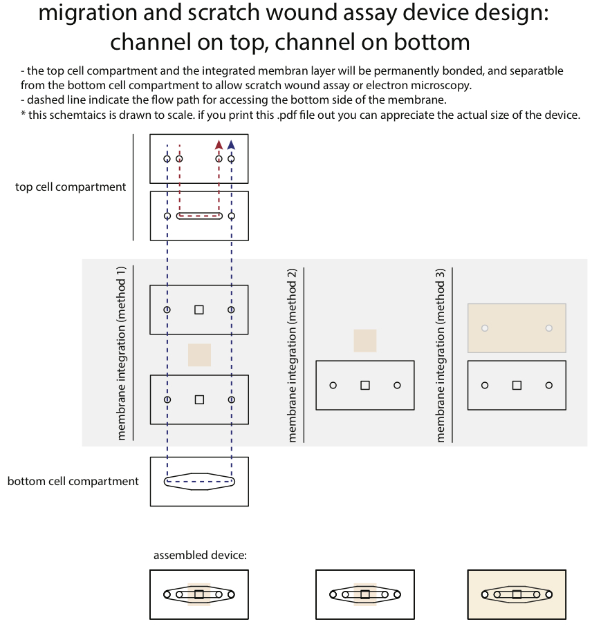
Microfludic Co-culture Device: