Neutrophil Migration and Endothelial Permeability – Closing remarks
So as most of you know by now, I have been working on developing microfluidic systems to study the leukocyte-endothelial interactions in vitro. I have developed systems that can facilitate the basic biological assays involving neutrophil migration and live-imaging. In this blog, I am presenting the latest (and the last) results on the effects of neutrophil migration on vascular permeability. To do so, I am using 2 different methods-electrical and chemical. Electrical methods rely on impedance spectroscopy, and the chemical methods involve monitoring of FITC-dextran diffusion across the endothelial monolayer as the neutrophils migrate from apical to basal chambers.
First, the impedance spectroscopy. I obtain the impedance at the frequency of 1000 Hz for 20 mins with one scan every ~60 seconds. The
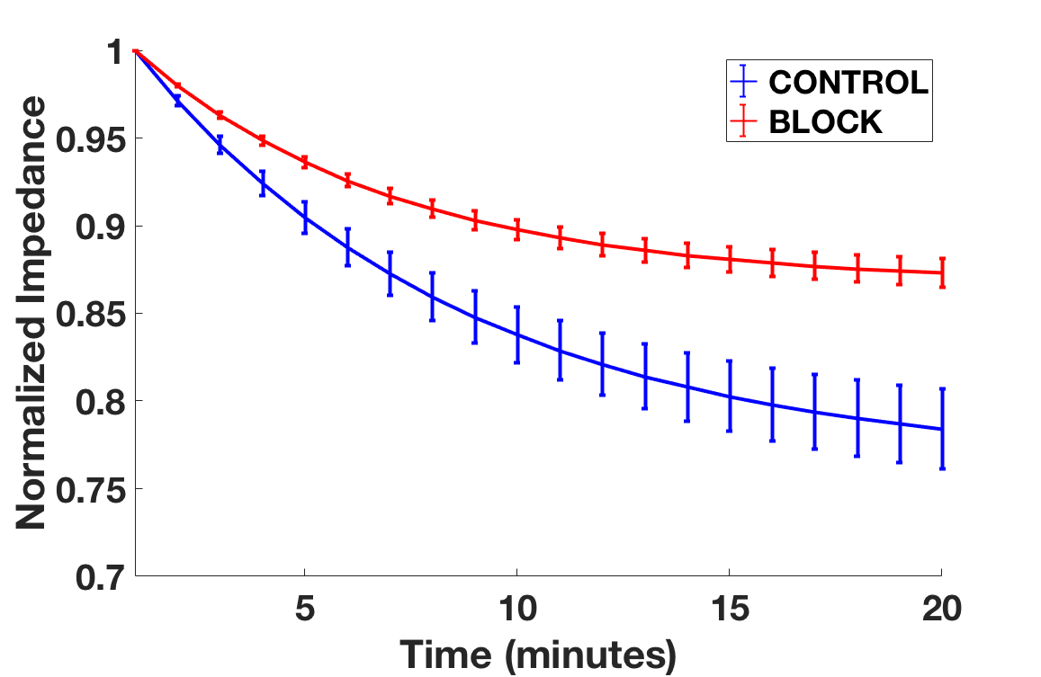
Raw data is fit using exponential decay, n=3 for both the cases. As we can see the decay in impedance is ~20-24% for untreated PMN, and the loss is about ~10-12% in presence of beta-1 block. This implies that preventing subendothelial (basement) migration of PMN can have some positive effects in controlling ionic permeability.
Next, I moved to chemical methods. As described in the Racquel’s blog, I am adding 10 kDa FITC-dextran in the top side and imaging the recovery in the bottom side for 20 mins, with images captured every single minute. Using this setup, I can get a better temporal resolution. Following is the raw fluorescence intensity data for the same treatment conditions.
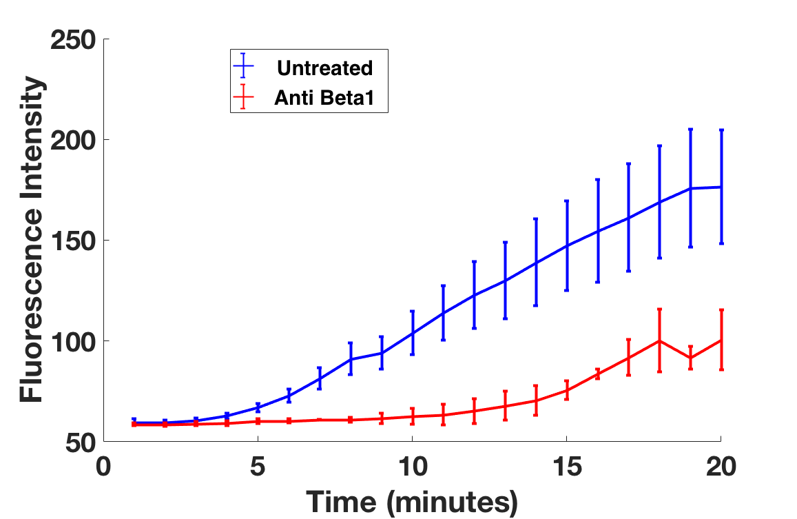
The rise in intensity is almost 2x more for untreated neutrophils (blue curve, n=5) as compared to beta-1 blocked cells (red curve, n=2).
Thus, both impedance and fluorescence data indicate that uninhibited neutrophils can migrate farther and cause enhanced leakage leading to increased net endothelial permeability. To further dissect this phenomenon, we decided to employ electron microscopy. In the past, we have used SEM to study the localization of PMN in the collagen gels during their transmigration process. We repeated the same assay but now with the
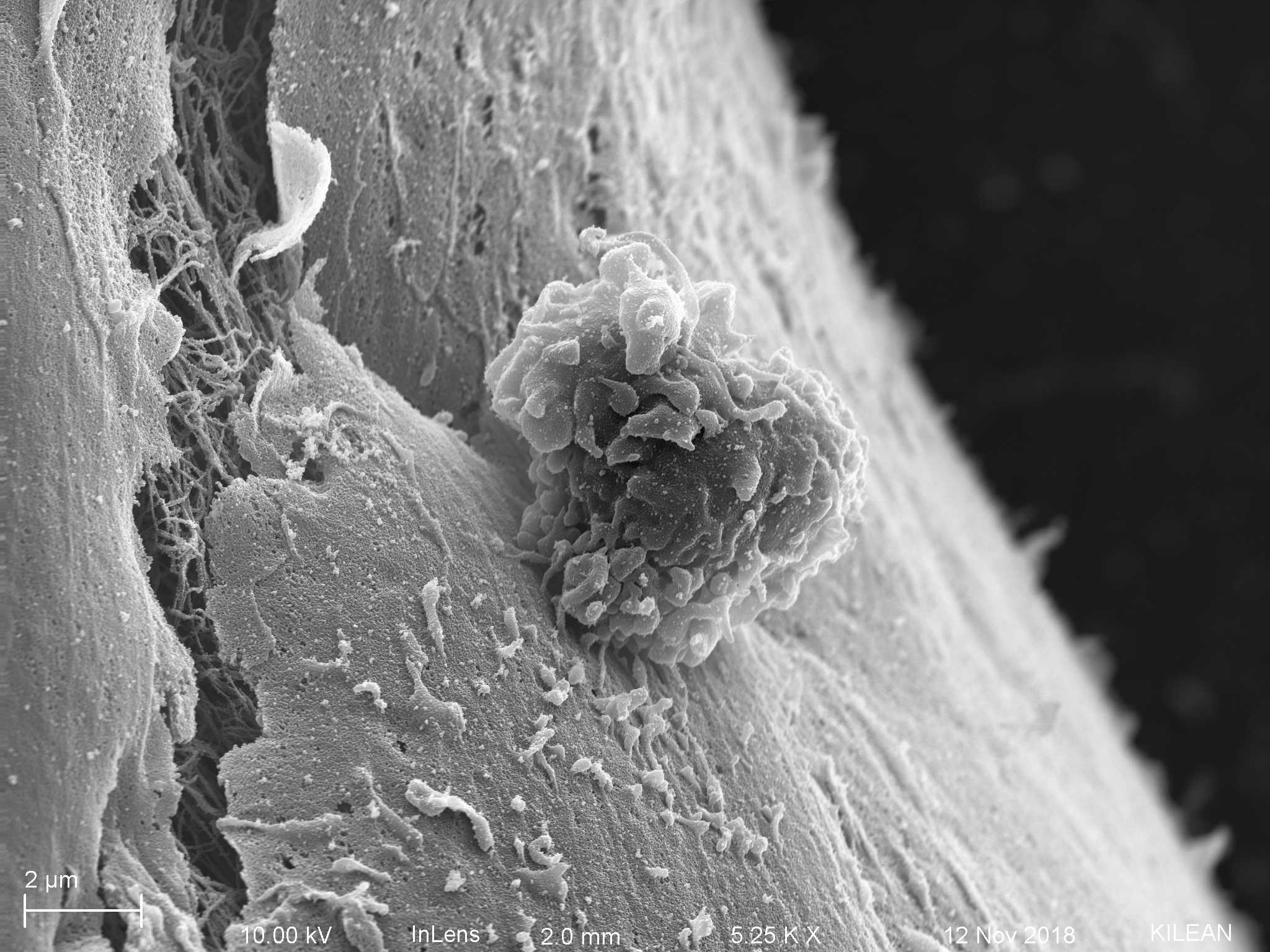

In comparison, this is the image we obtained last summer when there was no antibody block.
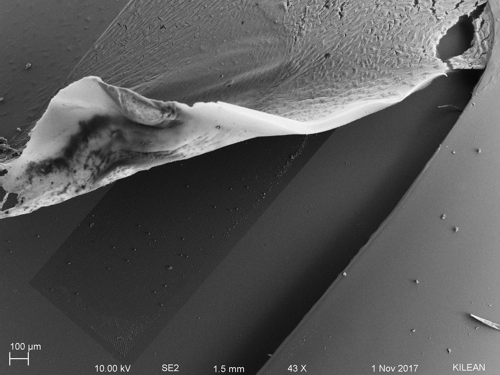
Although the comparison between the above 2 images is not overwhelming or quantitative, it does show the inhibitory phenotype of PMNs in the presence of beta-1 blocking antibody. This (preliminarily) validates the hypothesis that disruption of basement membrane can be responsible for increasing the net endothelial permeability, and preventing/trapping PMN in the subendothelial space can improve the barrier function.
Adios!