Thinkin’ Secretion in the µSiM
One of the many contributions Alec S. made to our group is to help us appreciate the importance of the apicobasal (A/B) polarity of endothelial cells. The effects of A/B polarity were demonstrated in his paper: Integrative Biology (2020), 12(11),275–289 where he showed that stimulation of ECs with TNFα from the abluminal chamber caused an even secretion of IL-8 in both directions, while stimulation from the luminal chamber caused IL-8 secretion primarily back into the luminal chamber. Given the chemoattractant role of IL-8, it was no surprise to find that luminal stimulation caused PMNs to remain in the top chamber, but it was a bit of a surprise to see that with abluminal stimulation, the elevation of IL-8 but with no apparent gradient, still promoted transmigration. In cartoon form, the results are summarized as …

The key data (derived from ELISAs and hand-counted PMN transmigration using live cell, phase contrast imaging) are summarized as …
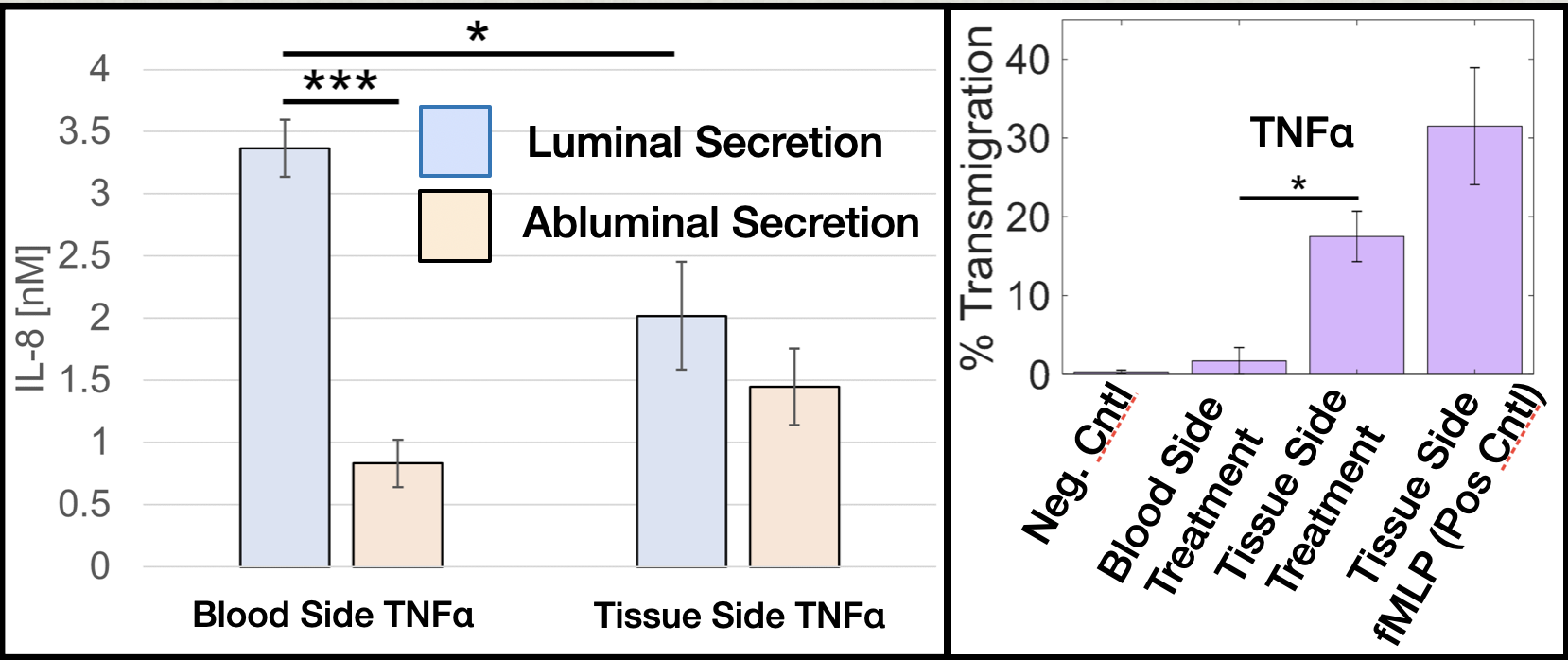
[Apologies for the proliferation of names for the ‘top’ and ‘bottom’ side of Alec’s devices (not the modern µSiM): We use: 1) luminal vs. abluminal (blood vessel perspective); 2) apical vs. basal (EC perspective); and 3) blood-side vs tissue-side (tissue perspective), seemingly interchangeably. Although we tried to pay attention to the context of our writing when making these choices in the paper. I am finding that for a more general scientific audience, tissue-side vs. blood side seems to be the most descriptive of these.]
An important issue was raised by Howard Su in one of our R61 small group meetings that relates to studies like this in the µSiM (Alec used handmade devices with a different geometry – not the modern modular µSiM). Howard pointed out that there are many more cells secreting into the luminal well than into the abluminal channel. Now its also true that the well volume is 10X larger than the backside volume, but it turns out this is not quite enough to lead to even concentrations in the case of uniform secretions in either direction. The key geometry is shown here …

And the key math is shown here … 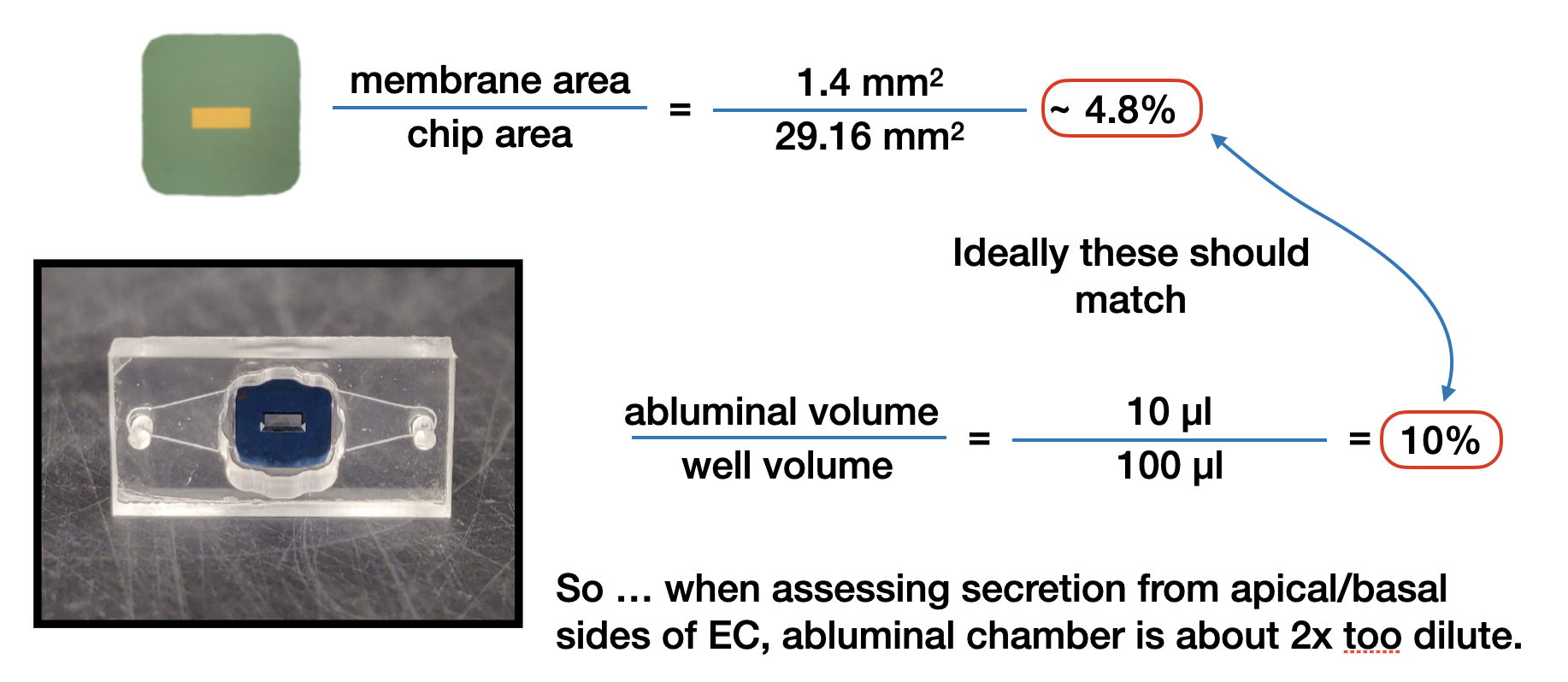 As for remedies, we’ve thought of a few …
- One is to add more volume to the well so that the apical secretion is diluted more. Unfortunately the max volume is 115 µL, so this would only reduce the error a little.
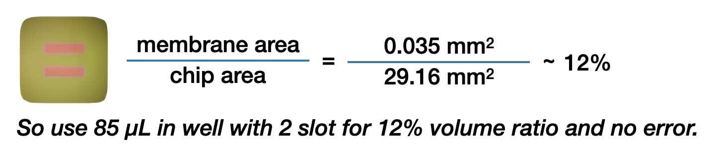
- It turns out our two slot membranes appears to be a great choice for straightforward interpretation of a/b secretion experiments.
- And finally there is Molly’s idea of just reporting the data on a per cell basis. In other words normalize both the well and abluminal channel concentrations by the total chip and membrane areas, respectively.