Depleted Serum Separation Attempt
I’m currently trying to compare separations between different complex protein mixture. This was my first crack at depleted human serum. This mixture has been processed so that the most common blood proteins (such as albumin and immunoglobulins) have been removed. All three trials were performed using membranes from w338 in diffusion mode. 338 has a fairly high porosity and large pores.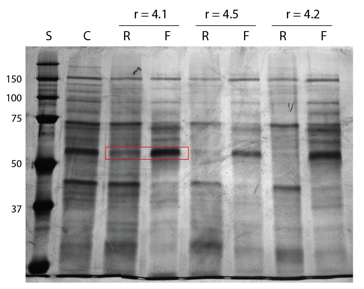
When you compare the retentates (R) and filtrates (F), you can see that no real separation has taken place in these trials. The membranes were selected so that there were no pinholes, but it may be possible that there were tears or that the pore size distribution was too large.
I outlined a set of bands that curiously is enriched in the filtrate. This protein diffused through the membrane much better than any other protein in this experiment in all three trials. We sometimes see a band that won’t pass as easily into the filtrate, and our arguement there is that it’s part of a much larger complex. I’m not sure how to explain why we see the reverse here. Maybe shape is the determining factor? Electrostatics shouldn’t come into play in a 1x PBS solution.
Another question for the proteomics center. What is this protein?
I forget how the dilution works out here. If the concentration of a molecule is the same in the retentate and the filtrate, the filtrate band will look much darker, because the retentate is diluted before running the gel, right? This compensates for the intrinsic dilution of the filtrate in the resevoir, right? Or are they just run straight now?
Since enrichment of the filtrate is impossible, I’m wondering if the dark bands are just nearing equilibrium. It’s also curious that some high MW bands are the same intensity…