Small Animal Model Dialysis: Nonporous and Microporous Membranes
On Wednesday Feb. 12 and Wednesday Feb. 19, limited exposure dialysis was performed on rats.
In the first experiment, blood was pumped with the peristaltic blood pump through a two-chip dialysis device in an acrylic fixture. The membranes were non-porous silicon nitride so no dialysate was included in the experiment. Blood was successfully pumped through the device with each chip receiving blood flow. Bubbles were observed to collect in the channels. The source of the bubbles is as of now unknown. They could come from a loose or cracked connection in a needle or stop cock. The blood was pumped through the device from the femoral artery to the femoral vein as in previous experiments. I’m sure we can locate the source of the bubbles but what troubles me more is the fact that they collect in the channels.

On the 19th. We repeated the experiment using 120 nm SiN with 8-µm pores. we first hooked up the Blood Pressure transducer to the carotid artery catheter to monitor the health of the animal. We are still having issues getting blood pressure readings in the range we expect. BP was 119/109, there should be a 20 mmHg difference between these two values. The pulse readings are well within expected values. HR = 310 bpm. While monitoring the dialysis device was hooked into the vasculature, first to the artery, to fill the system with blood and remove any bubbles from the saline which was preloaded in the dialyzer. The fluidic circuit was then completed by hooking the output of the dialyzer to the femoral vein catheter. Without assistance of the blood pump the pressure of the vasculature pushed blood through the dialyzer. The dialysate (saline) bag was hung and connected to the basal side of the membranes. At this time, blood was observed to be filling the output of the dialysate side. We then clamped off the dialysate to stop the flow. The blood pump was started and we observed the animal for 10 minutes. The blood pressure and HR were essentially unchanged. This is an important initial test to verify that the system does not cause the animal distress.

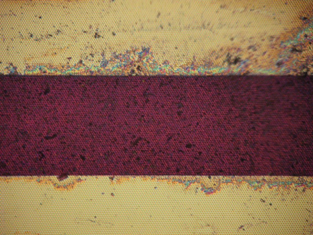
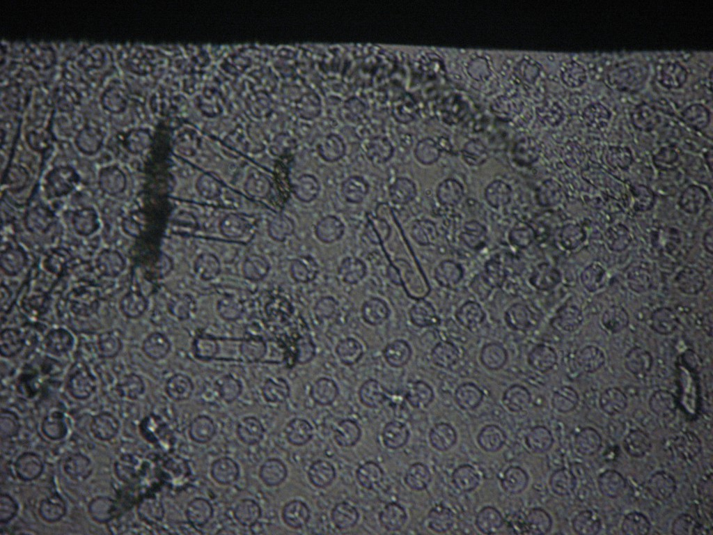
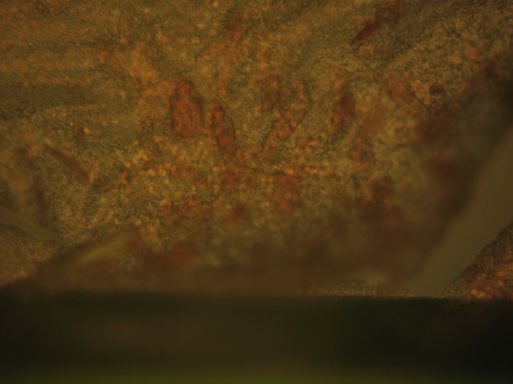
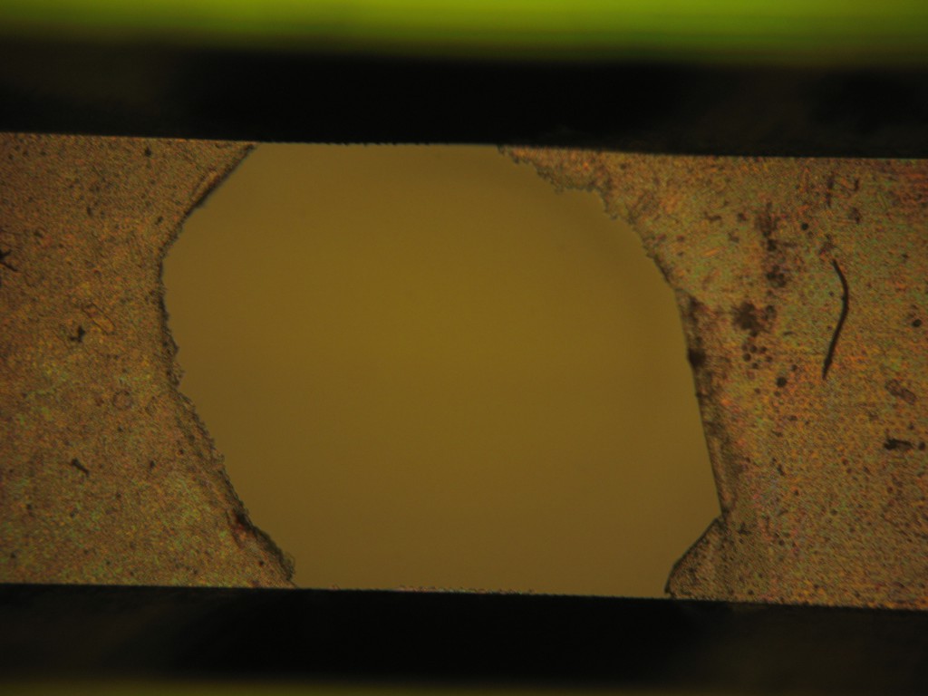
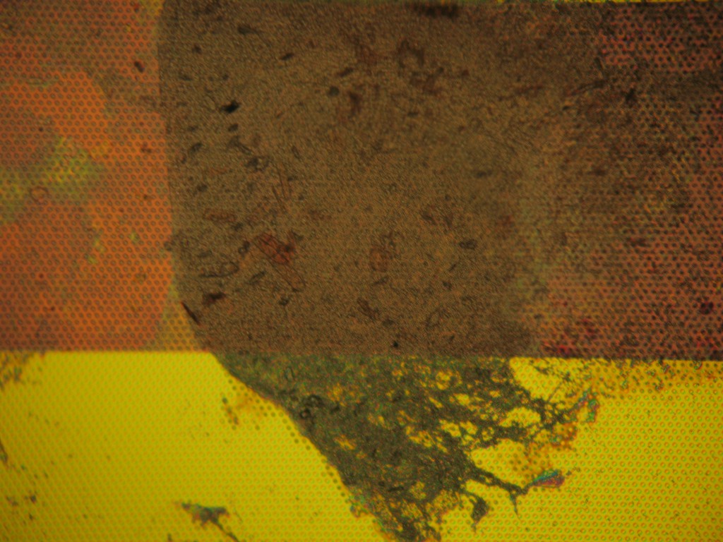
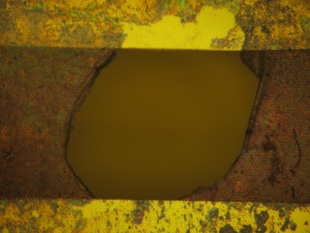
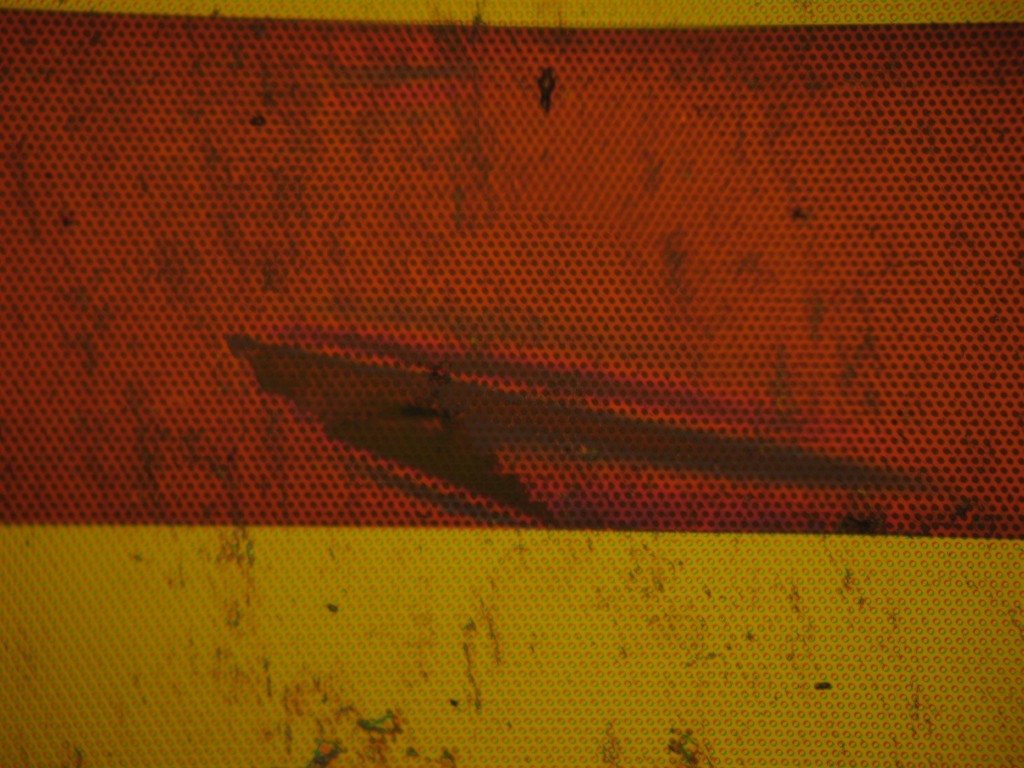
Post-mortem: The membranes were removed from the dialyzer and examined. One of the devices was entirely intact with no observable breaks in the membrane windows. The other membrane, however, had a large blowout (200 µm to 300 µm) across the channel and a couple small rips.
Of course the 8-µm pores wouldn’t have kept too much from the dialysate anyway.


Very interesting first step.
Any idea when the membranes broke? It does not seem like any of the pressures involved here could burst a membrane, correct?
Thanks!
The membranes were intact after assembly and flushing with alcohol and saline solution. They could have broken during transport (across the street), installation, or operation. I need remember my eye loupe to examine them during the procedure.
The pressure from pumping is minimal, < 2 psi in theory. More pressure may be applied by mishandling the tubing, which is often the failure mode for other membrane devices used in our lab. More bench top work would be helpful for this specific format.
Our membranes in fluid are extremely sensitive to impulse forces, like setting a hard device against a hard table. I suppose the lines could also create a pressure impulse, but since these tubes are soft, generating a substantial impulse would probably require a more substantial impact. I would either transport them dry, or wrap them in something soft. Even sticking some felt or rubber feet on the device may make it more resistant to impact forces….