Elder Lab Staining Airborne Microplastics
Elder Lab Staining Airborne Microplastics
Materials
Biological Safety Cabinet (BSC)
Side arm flask, 150-1000 mL
Small pin Fritted glass filter
Vacuum and tubing
0.5 µm Sepcon Filters
1.7 mL microcentrifuge tubes
Microcentrifuge
ddH2O (Barnstead 18 MΩ)
100% isopropanol (IPA)
100% Ethanol (EtOH)
70% Ethanol (EtOH)
0.4% Gibco Trypan Blue (TB) – 1 year shelf life
99.99% MCE Nile Red (NR) Powder
20 µL and 1 mL pipettors
Amber Glass vials for stain storage
250°C Oven
Gloves (nitrile)
Parafilm
100% Cotton Lab Coat -No Plastic Fibers
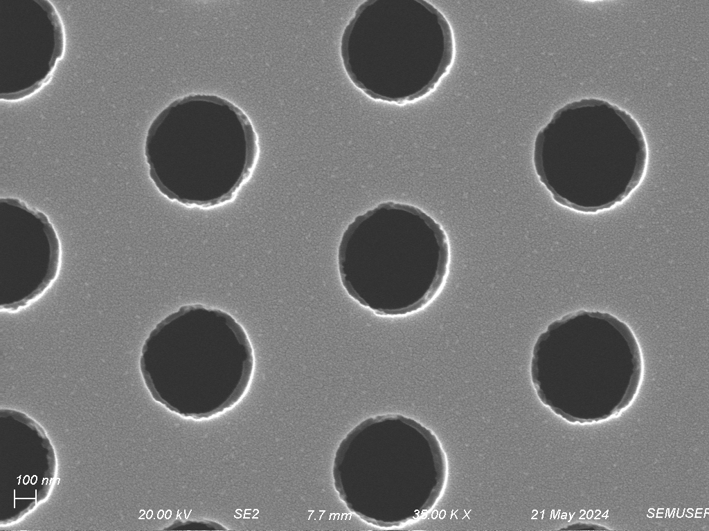
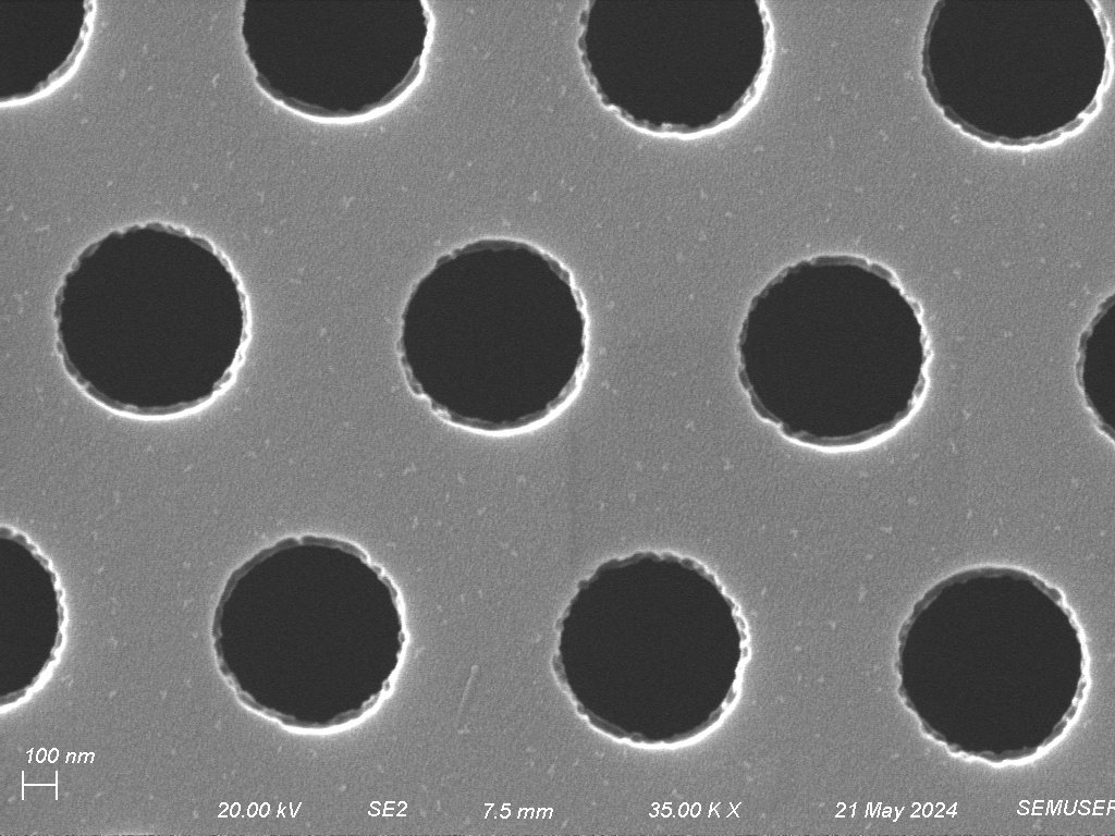
SiMPore Filters with nano/microparticles
Tweezers cleaned with IPA to manipulate the SiMPore filter from container to the fritted glass filter
Lionheart Plate Reader – Ben Miller Lab
All glassware is to be cleaned per SOP MP-4 Glass and Reagent Preparation
Procedure
- Sample Collection
See “GilanSamplingRev1_2.docx” for sampling procedure
- Stain Preparation
Wash hands and put on lab coat and gloves. Within the BSC, spray everything with 70% EtOH and wipe using Kimwipes. Nile red is prepared by mixing ~50 mg in 45 mL of 100% EtOH in a centrifuge tube. About 10 uL of this is diluted with EtOH to 1µg/mL to yield 10 mL, thoroughly vortexing ~30s before and after adding the 10 uL. Depending on the initial mass dispensed, calculate the remaining volume needed from the stock tube to dilute the second tube to achieve 1 µg/mL. In an amber glass vial, add 1 mL of 1 µg/mL NR, then dilute with 4 mL of ddH2O. Into an amber glass vial cleaned per SOP MP-4, use a Sepcon to filter the 5 mL of working concentration NR (0.2 ug/mL), and keep up to 30 days.
Example:
50 mg NR Powder/45mL EtOH = 1.111 µg/mL
(x mL)(1.111 µg/mL) = (10 mL)(1 µg/mL)
X mL = 0.009 mL = 9 µL
(9 uL)(1.111 µg/mL NR) + (9.991 mL EtOH) = 1 µg/mL in EtOH
(1 mL)( 1 µg/mL NR) + (4mL Sepcon filtered ddH2O) = 0.2 µg/mL NR in 20% EtOH
Cf = 0.2 µg/mL
Vf = 10 mL
4 mL of commercially prepared 0.4% Trypan Blue from Gibco is diluted to 0.04% in 0.9% Saline and centrifuged through a Sepcon filter into an amber vial.
- Staining Procedure
Wash hands and put on lab coat and gloves. Within the BSC, spray everything with at least 70% EtOH and wipe using Kimwipes. Remove the parafilm sealing the glass Petri dish containing the SiMPore filter using tweezers. Place the filter on the fritted glass filter facing up and the troughs pointing down. You can identify the bottom by how the filter is seated in the plastic housing unit. Do not touch the filter with the pipette tip. Try using your other hand as a fulcrum to reduce motion.
For Nile Red, apply 60 µL in 10 µL increments in a sweeping motion across each of the 3 windows with the vacuum on (20 uL per window). The vacuum is left of for at least 1 minute to let dry before turning off and returning the filter to its Petri dish for imaging.
If desired, apply a 20 µL drop of 0.04% Trypan Blue to the filter and let soak ~1 min after staining with NR. Turn on the vacuum and wash 2 times with 250 µL of ddH2O being careful not to let water touch the black disc and fall through filter. After the washes are completed, turn off the vacuum.
- Imaging
See SOP “Using the Nikon Epifluorescent Microscope in Goergen Hall B109” OR “SOP Lionheart FX Microscope – Rm 5-5144.docx”
- Clean up
Put the vacuum tube away. Disassemble the side arm flask and fritted glass filter and wash with Dawn/Green Clean/ etc. Rinse glassware with tap water followed by a 2-5% Alconox rinse. Rinse 3 times with 0.22 µm Barnstead ddH2O and fill with IPA. Sonicate the pieces for 3-5 minutes and cap the pieces with aluminum foil as they are pulled out of the sonicator and bake in oven at 250°C for 6-12 hours. Reassemble the pieces and cover with aluminum until next use.
- Interpretation
See SOP “Image J Airborne MPs” and “Lionheart Meta Data”. Blue stained objects are cellulosic, e.g. cotton. Nile Red positive objects are very likely to be plastics, but composition needs to be confirmed by other specific techniques.