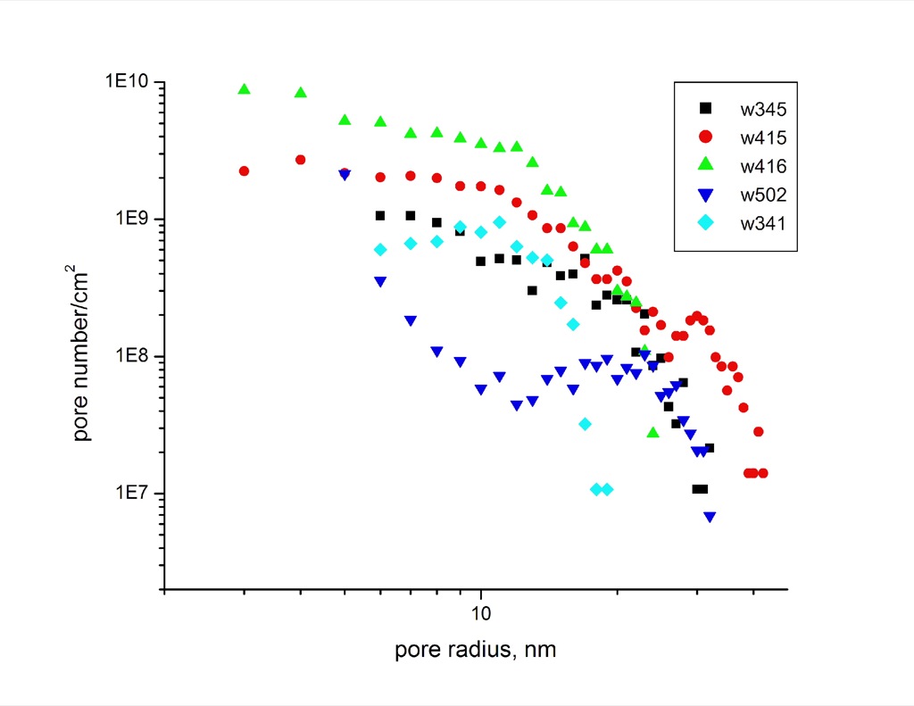
-2 Power law for density vs. pore size
I realized last week that I had made a pretty substantial mistake with my quantification which involved my mass standards. Half way through running the gels for the previous study I had accidentally started making my mass standards at a higher concentration but counting them in the quantification as their initial value. The bottom line…
An initial experiment for membrane deflection characterization using an optical profiler (Veeco Wyco) has been performed. Below are some of the images taken with the tool: (a image was taken at each 0.5 psi interval): Once we get a pressure regulator we will be able to generate numerical data of membrane strength/deflection down to 5.0…
Summary After success with the development and execution of the μSiM-CA (Canalicular Array; see Masters et al., Nanomedicine: Nanotechnology, Biology, and Medicine 2019), Jim wanted to develop a platform to answer some more biophysics focused questions. In order to facilitate these studies, the platform would ideally be configured to allow for live imaging of staph…
Previously we have studied protein adsorption to pnc-Si membranes by TEM and fluorescence. Neither of these methods will work with polymer membranes. Polymer membranes are too thick to view using TEM. Polymer membranes also autofluoresce creating a lot of noise while using our fluorescence method. Here I outline a new method of studying adsorption to…
For the first time, we did time-lapse phase microscopy (1 frame / 5 seconds) looking at endothelial cells through the ITO in the static BBB device (device A bottom left well) and measuring the TEER concurrently. Here’s the video, just shy of 15 minutes of recording. (reporting a low resistance: 5-10 ohms, shorted). TEER Test…