Contact Angle Study
According to recent cell adhesion studies on aminosilanized pncSi, no significant difference between adherence to the silanized and non-silanized surfaces was found. This could have been due to the adsorption of protein from the cell growth media onto the surface nullifying the effect due to silanization which is supposed to make it hydrophobic.
‘If silane treatment is making the surfaces hydrophobic, it should impact cell adhesion and spreading, but it is likely that proteins coat the surface during spreading and perhaps they pacify the surface and facilitate attachment just the same.’
So contact angles were measured for non-silanized and silanized samples with & w/o incubation in EGM (+FBS) (serum supplemented Endothelial growth media). All samples were from W684 (no RTP) with intact slits. After being silanized, the samples were incubated in preincubated media for ~25minutes. Then they were rinsed gently with HBSS and left to dry on benchtop.
4 ul dH2O drop was put on each surface and imaged carefully keeping the surface horizontal and in same plane as the camera focus.
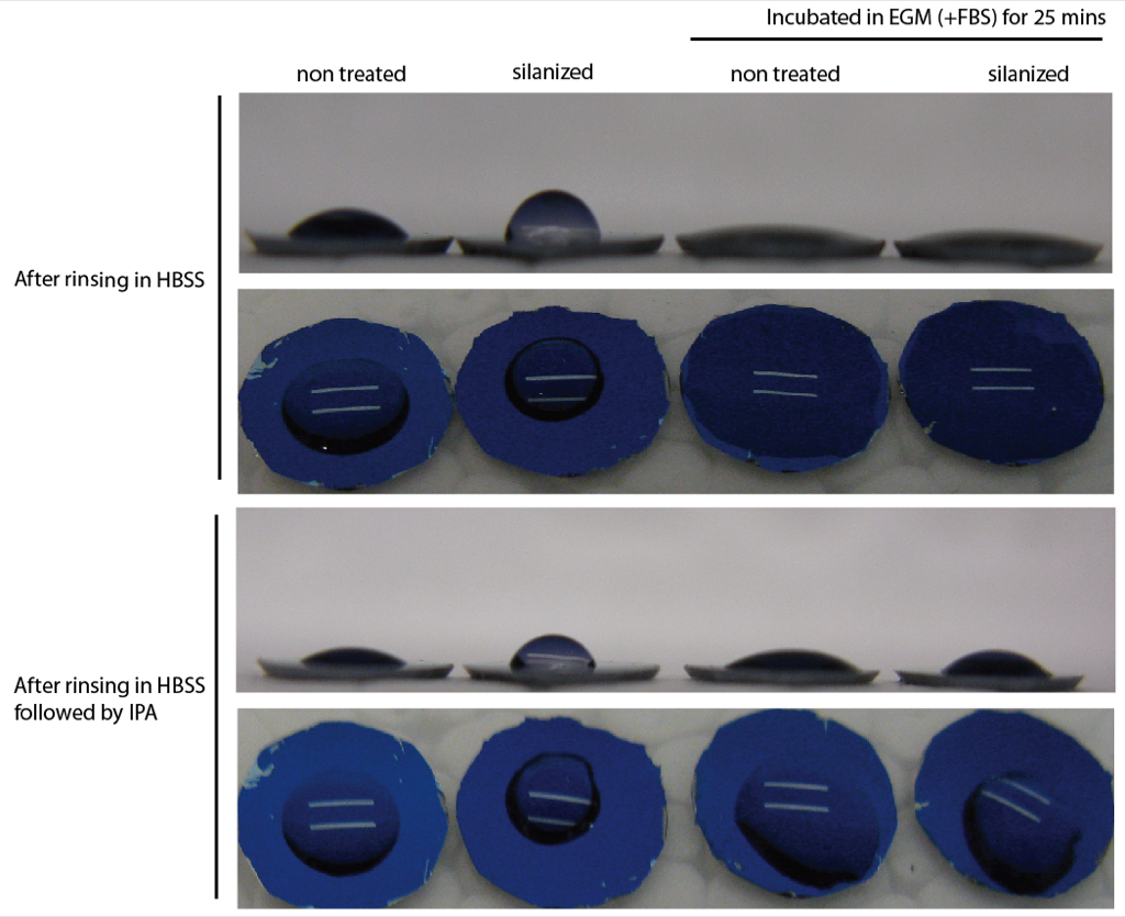
In above figure, the first set represents samples which were rinsed in HBSS after incubating in media. They were left to dry on benchtop and then water drop was put on it. Since the drop went almost flat on the surface, I wasn’t sure if the surface had completely dried. So I rinsed the samples again in IPA which helped drying it completely, and then second set of images were taken. This time the drop didn’t go flat, although formed fairly low contact angle (same as non silanized sample).
Hence, the proteins from the serum supplemented media coat the silanized surface and pacify its hydrophobicity allowing cell adhesion. I had multiple silanized samples, all of them gave similar contact angles, showing that this is repeatable.
The contact angles measured from the above images using Image J are approximately:
| Non silanized | Silanized |
Non silanized
(in media) |
Silanized
(in media) |
|
| HBSS rinse | 35-40 | 70-75 | 10 | 15 |
| HBSS + IPA rinse | 35-40 | 70-75 | 25-30 | 35-40 |
Nicely done. I’m not sold on the cell adhesion data just yet because of the problems with cell morphology. And we need to do spreading and growth as well. There is a probably a negative consequence here for cells growing in defined media (rather than FBS) because the protein levels are much lower. Anyway, lots to do to wrap this up, but its all very straightforward and we can organize starting Tuesday.
The contact angle for the silanized samples is a little high, but if it gets coated with protein, it probably doesn’t matter for this work. I think a good APTES monolayer should have a contact angle around 50 deg. What you likely have is more than a monolayer or perhaps a semi-inverted monolayer where the hydrophobic silane groups are more common than amines on the exposed surface. Once the YES system arrives, I suspect the contact angle will go down. You may want to try a buffer rinse AFTER baking, to remove the non-covalently attached silanes, if it matters at all. This is likely what happens when you apply the media, in addition to the protein adsorbing.
So why does cell adhesion in silanized samples seem to be lower than in unsilanized samples? This data indicates that there shouldn’t be much of a difference.
I don’t think there is a significant difference. In the first trial of cell adhesion it looked similar, but in the second trial in particular, the difference could be seen because the silanized samples had broken slits leading to loss of cells.