"Long-term" Cell Culture on pnc-Si transwells
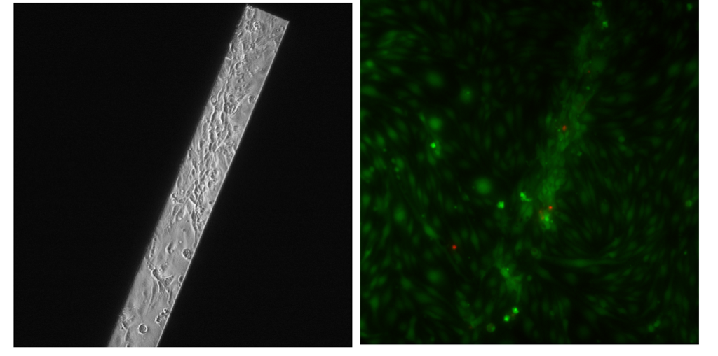
Along with the PET data I just posted, I also examined cells on pnc-Si transwells. The following images were taken at 20X (through the bottom of a 24-well plate), and the cells are passage 8 bEnd3. They were seeded on the membrane side @ 100000 cells/cm2, and then the transwells were flipped for growth. Images were taken 7 days after seeding. Staining was with Live/Dead. pnc-Si was SC126, RTP’ed.
The left panel is a phase contrast image of 1 slit – cells look pretty good but there seem to be really densely packed cells or maybe even a multilayer of cells over the membrane. The Live/Dead panel really brings this out. There is a confluent monolayer on the supported membrane but really dense packing of cells over the membrane. In a couple of these densely packed areas, there are dead cells.
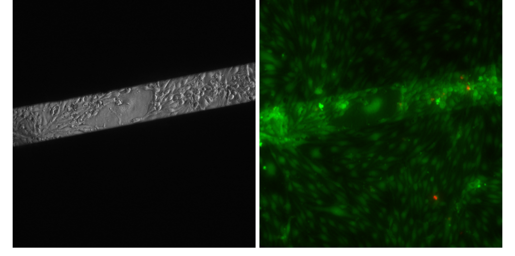
 A different pnc-Si sample showed the same thing. The cells seem more dense and rounded over the membrane compared to the supported area. It seems like there might be a multilayer of cells toward the left off the membrane and more rounded cells on the right part of the membrane. Here, you can see a non-endothelial cell over the membrane in the middle of the membrane. Again, there are some dead cells around the cell ‘clumps’.
A 3rd sample shows the same thing, except the cells over the membrane are particularly bad-looking. It looks like there is a monolayer of cells extending over the membrane and then clumps of rounded cells either above or below the monolayer.
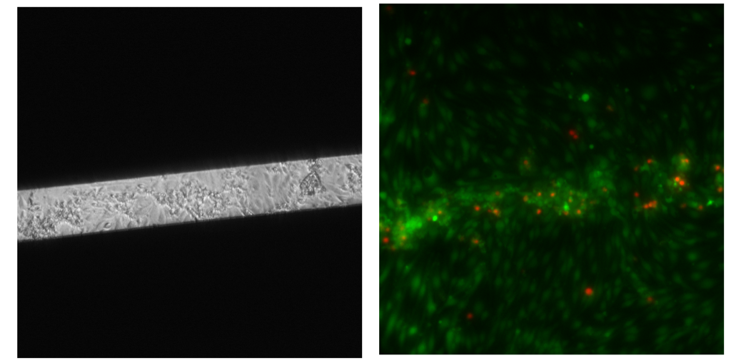 Obviously, this is a problem. What’s nice is that the dead cells correspond to the darker, round, “clearly not endothelial morphology” cells in phase. This means I can flag problem samples without having to do the Live/Dead stain.
Some ideas on why this is happening: 1. I’m plating at too high of a density, allowing (by chance) some dead/almost dead cells to adhere to the membrane (or a sagging membrane), where they hang out and get covered or pushed up out of the way of growing endothelial cells. 2. The cells are proliferating especially well over the membrane due to nutrient access from both top and bottom. 3. The cells are pulling on the loose (compared to supported) membrane, recognizing the different mechanical properties of the free-standing film and reacting to it.
Any other ideas?
This data is very interesting. Do you think the cells can sense the different mechanical properties of the membrane relative to the wafer? My first impression would be no, because the membrane probably seems quite tight to a 10um object. We have to apply a lot of pressure to deflect the membranes a few microns, so over the size of the cells, there would be little curvature. Do cells “touch” surfaces in a way that would be sensitive to nanoscale deflections? My vote would be some type of transport through the membrane, diffusive or convective that the cells don’t like.
There’s certainly a mechanical difference between supported and free-standing membrane – I think the cells might detect this (a 10um cell in the middle of the free-standing membrane) might sag the membrane slightly. If the membrane sags during cell seeding, cells could accumulate over the membrane (especially at the super high seeding density I typically use).
Nanoscale differences in topography could affect the cell adhesion/migration, but the topography on supported and free-standing membrane should be the same. So I’m hesitant to blame it on topography.
I like the idea of enhanced transport/flow through the membrane. Perhaps there is a slight water flux that slowly draws cells toward the free-standing membrane? As they congregate over the membrane, they restrict nutrient access to each other?
I’m going to try a lower seeding density to minimize initial cell aggregates.