Blood flow and chamber properties
Hi everyone!
I’m sifting through the mass of data I collected during my last month and am compiling posts to update everyone. This post shows the latest with the microfluidic chamber that Henry and I designed. In my first post regarding this device, I showed microparticle and blood flow over cultured endothelial cells (bEnd3). In the movie with blood flow, we saw cells blebbing on the membrane. I repeated these experiments with blood flow but no bEnd3 cells to see if the blood cells were reacting to pnc-Si.
In this movie, RBCs flow above the slit and some adhere to the membrane but their biconcave morphology doesn’t change during this 1-hour movie. Therefore, it seems like the blood cells do not bleb by themselves. This raises a couple of interesting questions: 1. Are the bEnd3 cells reacting to fluid flow and blebbing? 2. Does the presence of bEnd3 cells (or their secreted/adsorbed proteins) on 1 side of pnc-Si membranes cause RBCs/leukocytes to adhere to the other side of pnc-Si and activate? 3. Did something else in my previous prep cause leukocyte activation and that’s what we saw in the previous movies?
For those who haven’t seen these devices, here are a few images and properties.
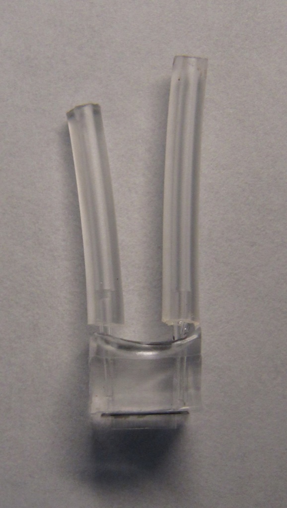
Side view shows the PDMS chamber with glass capillary tubes protruding from the top. Tygon tubing is connected to the capillary tubes for flow (from a syringe mounted on a syringe pump).
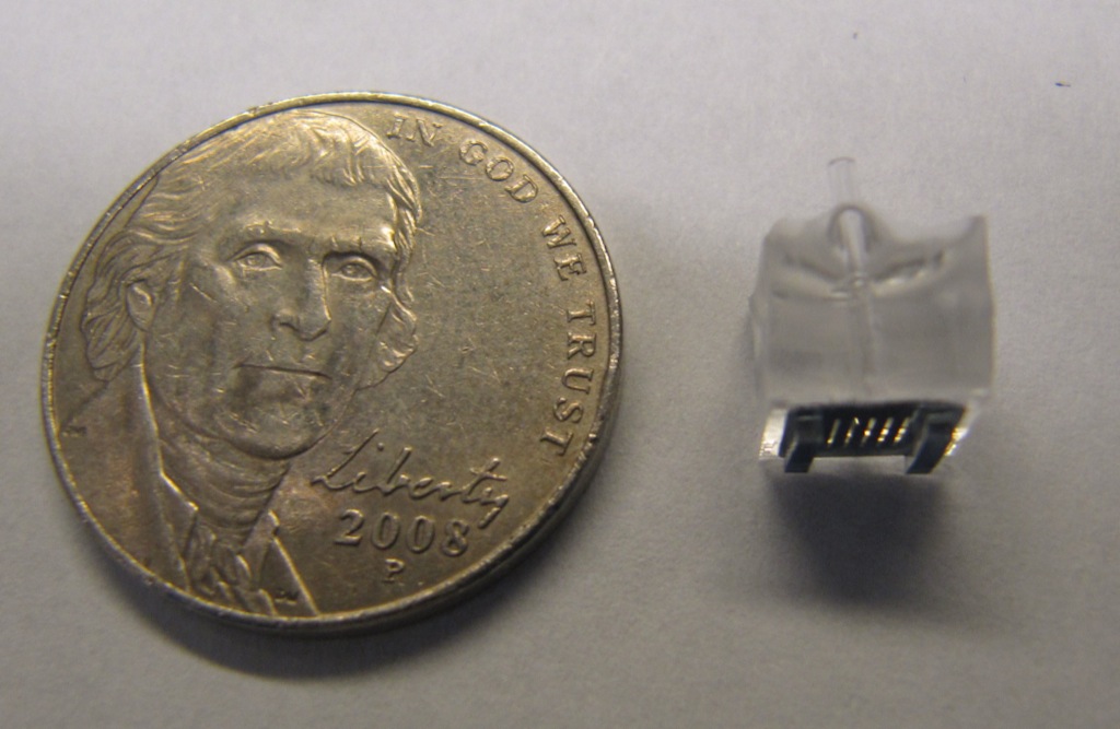
Bottom view shows the 5-slit square pnc-Si chip. There are 2 PDMS rectangular ‘legs’ that act as spacers so that the device can be placed on a surface without breaking the slits. (This is the side I cultured bEnd3 cells on for the last post).

A different side view shows the transparent slits and the PDMS ‘legs’:
This schematic shows that relevant heights and a little more detail on the chamber for flow – please correct me if I’m wrong, Henry.
