BBB V2.8 @ Vaccinex Adhesion Molecule Testing
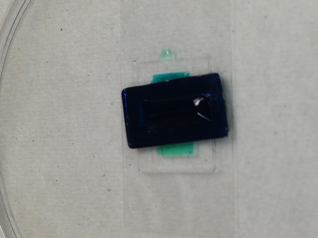
I created some quick open air devices to test the viability of different adhesion molecules. The basal well has a volume around 150 uL, and the apical well has a volume around 15 uL.
I made 6 devices in this format. On 5/14/13, Alan and I filled the basal chamber with PBS and then placed 20 uL of different adhesion molecules on the apical chamber. These coatings were incubated at 37C for 1hr. After incubation, the adhesion molecules were washed off with PBS. Cells were then plated with CSC complete media at a density of 0.5e6 cells/mL at 3:45pm.
- A – uncoated
- B – Collagen
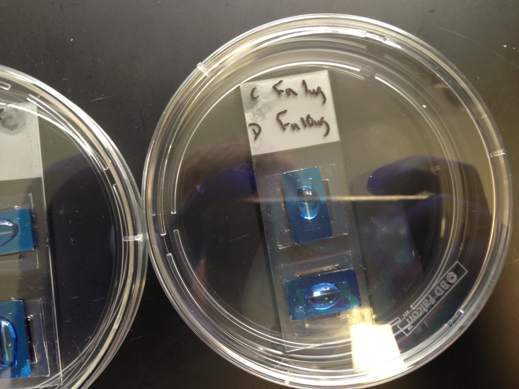
- C – Fibronectin 1x
- D – Fibronectin 10x
- E – “Adhesion Molecule” (believed to be some sort of gelatin)
- F – Gelatin


The surface is pretty hydrophobic. Alan manually dragged the fluid boluses across the flat side in an attempt to get an even coating. The cells were allowed to settle out of solution and reach the membrane for 45 minutes. Here are some pictures showing the seeding state of cells at this time point.

There are a couple of cells starting to spread out in the uncoated device, but not bad. Density is uneven.
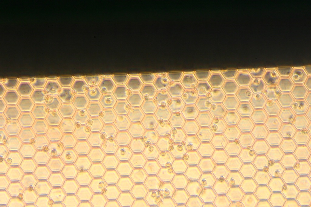
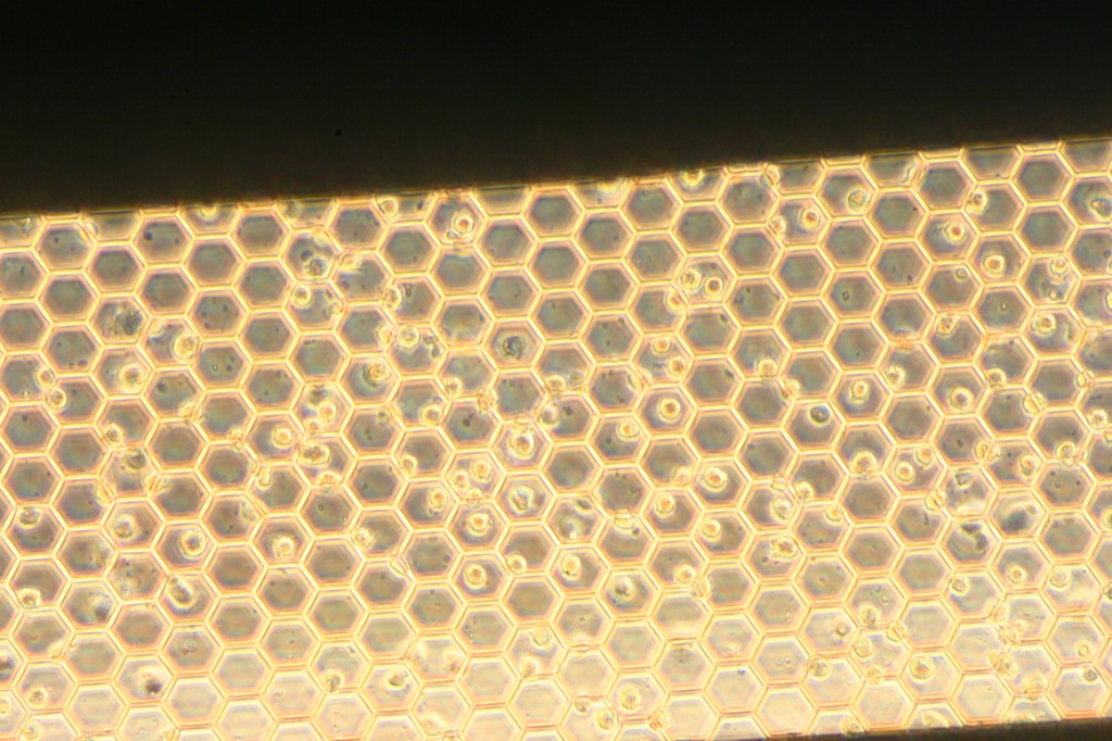
Quite a few cells starting to lay down here. Decent density across the entire membrane.

Few cells starting to lay down here. Much higher density in the middle.
Not so many cells laying down here.
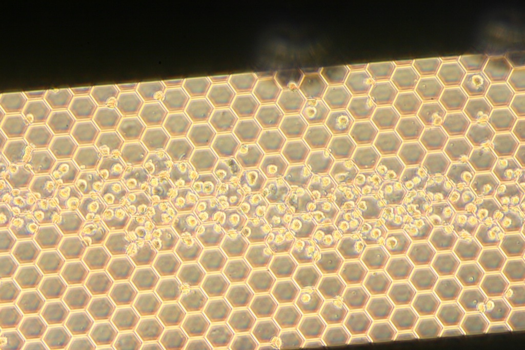
This is a very interesting distribution. We believe that flow from the apical chamber to the basal chamber is causing them to agglomerate. It also seems that the cells prefer to stick to each other compared to the membrane, which would explain the clumping of cells we saw in device #3.
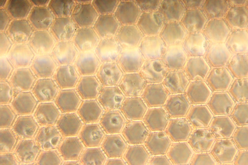
Here too, cells are beginning to lay down. We are optimistic that the gelatin, collagen and fibronectin treatments will cause the cells to form a monolayer.
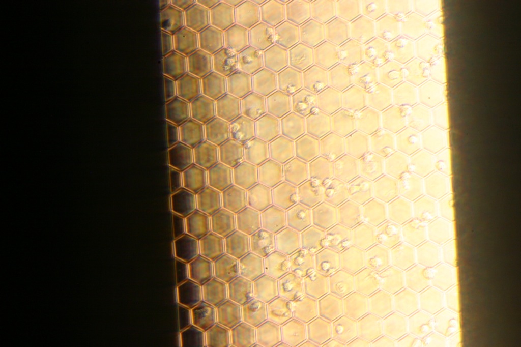
Update 05/15/13 – 22hrs since seeding
Here are the same samples after 22hrs. The media was changed in both wells and placed back into the incubator.

The cells here are loosely attached to the membrane. When we move the sample, the cells move with the media. There are a few cells that are laying down into a fried egg. According to Alan, they looked much healthier 6hrs earlier, checked in the morning.


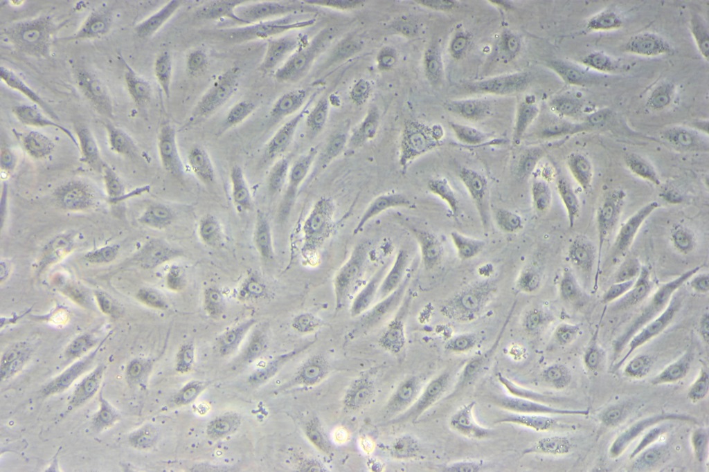
The fibronectin samples overall have the most spread out cells, and have a pretty good density after a day.
Few clumps and a few cells. Much like what we have been observing in the device.
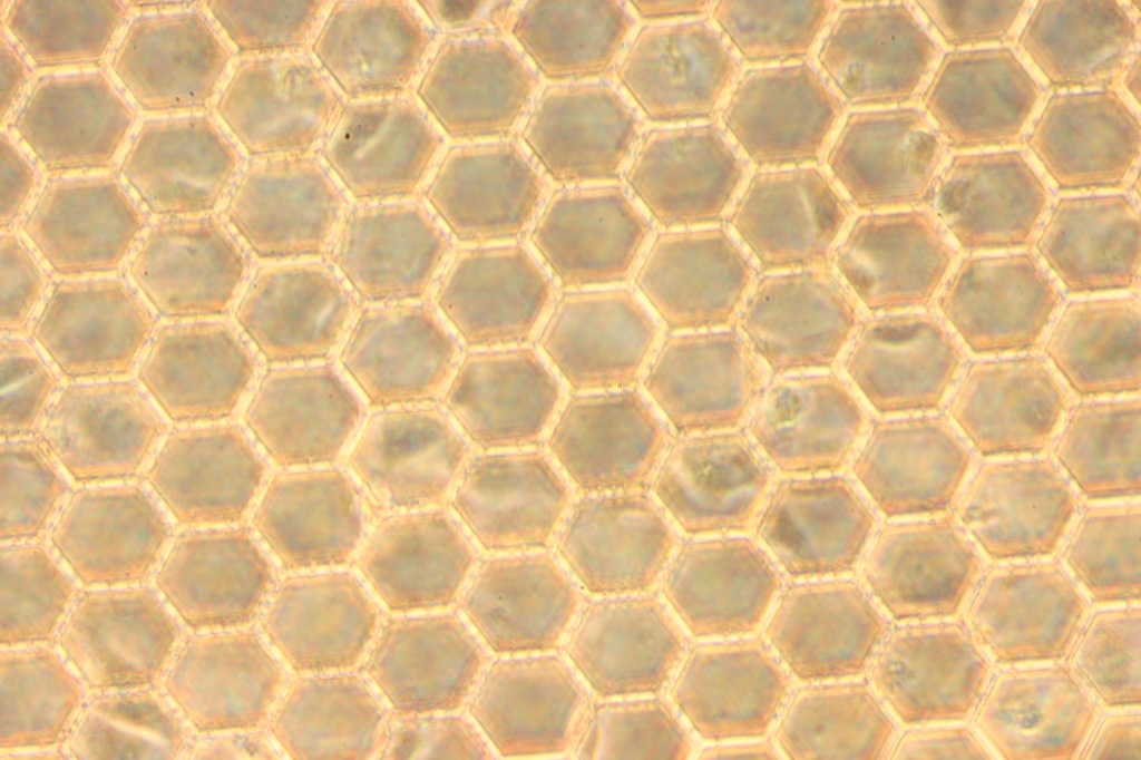
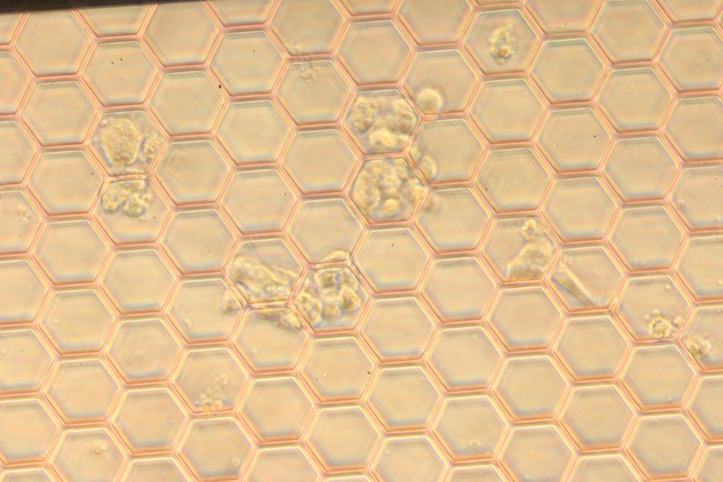
Gelatin looks pretty good, but the cells are loosely attached to the membrane. Shaking the sample causes the cells to shake along with the media, not the membrane. Dead cell parts are clearly evident. What are the little microbubbles forming on the hexes?
Update 5/16/13: 40hrs after seeding
Based on the results from today and the day before, we are selecting Fibronectin 10ug/ml as the adhesion molecule of choice. We will be incubating a device with the fibronectin, seed it, let the cells grow overnight, and then put shear flow on it tomorrow.
Should we plasma/ozone treat beforehand to address the hydrophobicity?