Fluorescence Focal Test – 300nm Microporous Oxide Membranes Do Not Wrinkle or Sag After Autoclaving
Method:
Oxide membranes in some conditions have been shown to wrinkle over time. To study this, three different varieties of SiO2 membranes were observed (1000C nonporous 120nm SiO2, 150W deposited microporous low-density 300nm SiO2, and 100W deposited microporous low-density300nm SiO2). Beads were placed on the top of each membrane and given ~24 hours to settle down. Images were taken in 10x to show either presence or lack of sagging.
Conclusion:
The results show that the 1000C annealed 120nm membranes have substantial wrinkling and bowing, while none of the new 300nm membranes displayed bowing to the degree of focal difference.
I had previously posted on the transparency of various substrates in regard to fluorescent beads, normalized to glass.
Adding to the study of beads on membranes, I actually looked at the wrinkling and ‘bowing’ that occurs over time, especially evident in the 1000C annealed oxide membrane. The one I used was a nonporous 120nm oxide membrane chip from wafer #1034. When dry, the membrane is extremely wrinkled in macroscopic appearance. However, when wet the membrane appears transparent:

The moisture doesn’t make the membrane more taught, however. I believe it just gives the membrane the opportunity to ‘bow’ downward, having more pressure on it from the water versus air.
When looking under the microscope in regular transmission microscopy, the same conditions (first is dry, second is wet) seem to be indistinguishable from each other. Images are representative of 1/4 of the membrane, the top-left corner specifically (scale bar is 100um). (The ‘waves’ are caused by the petri dish holding the device; the light gradient in the second image are likely an artifact of the water bolus, seen above).
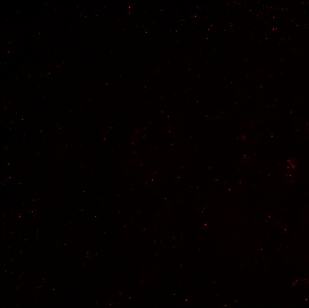
To observe any sort of bowing action that was occurring, beads were placed on the membrane and given about 24 hours to settle. Afterwards, images were taken at different focal planes to see any difference in the beads’ height (see below schematic).
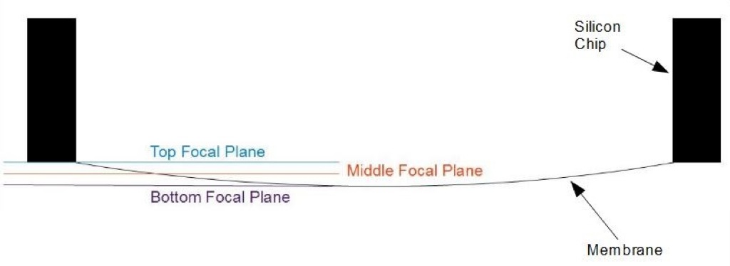
Beads were only placed on top and the membrane is nonporous, so none could have leaked through. When imaging, it appeared that the beads were in different focal planes, indicative of a ‘bowing’ of the membrane (all images 10x, scale bar is 100um). First image is the base focal plane, second is 25um below that, third is 48um below the first).
The following .gif file is a timelapse in fluorescence (file is b&w due to saving/conversion). The bottom left begins in focus, then focal plane is lowered to bring the top right into focus. The bottom corner is then focused on again; the focus is finally shifted down through the focal planes on the membrane into a state of being out-of-focus at the end.

As this timelapse was taken in fluorescence, any bolus on the top of the membrane would not have affected the emission light, and therefore this focal shift is indicative of a bowing of the membrane.
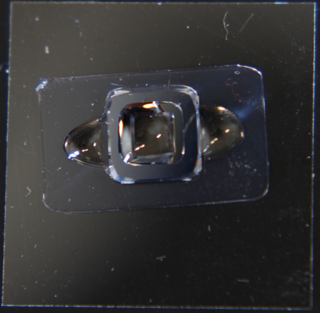
A similar experiment was repeated using the new oxide membranes. Four total were made into single-well CytoVus, all at lower pore density. Two from the 100W deposition and two from the 150W deposition were used, one of each being autoclaved beforehand to see if that caused any additional wrinkling or bowing.
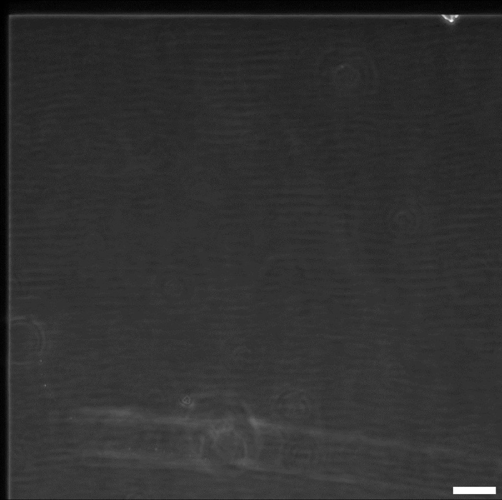
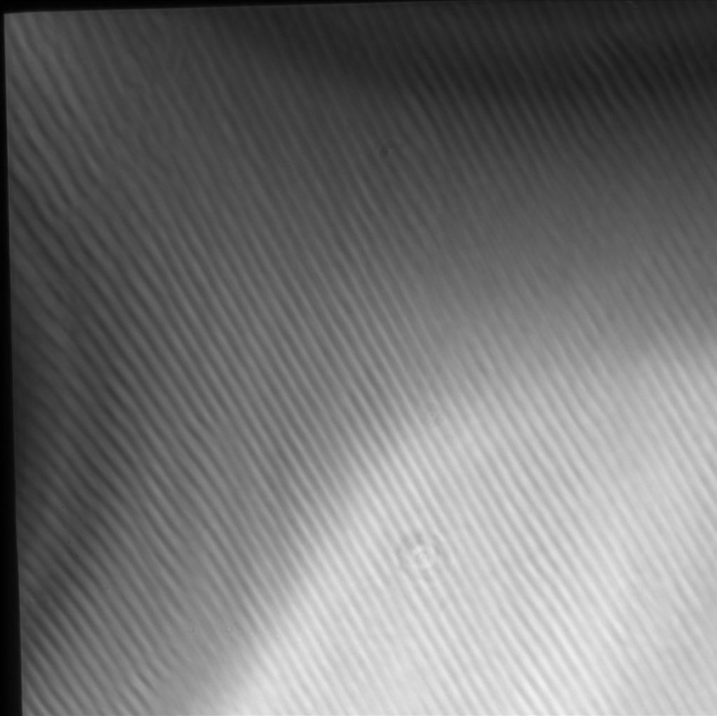
It seems that all membranes were fairly flat, and had little to no shifting in focal length from corner to center of membrane. One representative image from each was taken (all same magnification & orientation as previous images):
100W, Not Autoclaved
150W, Not Autoclaved
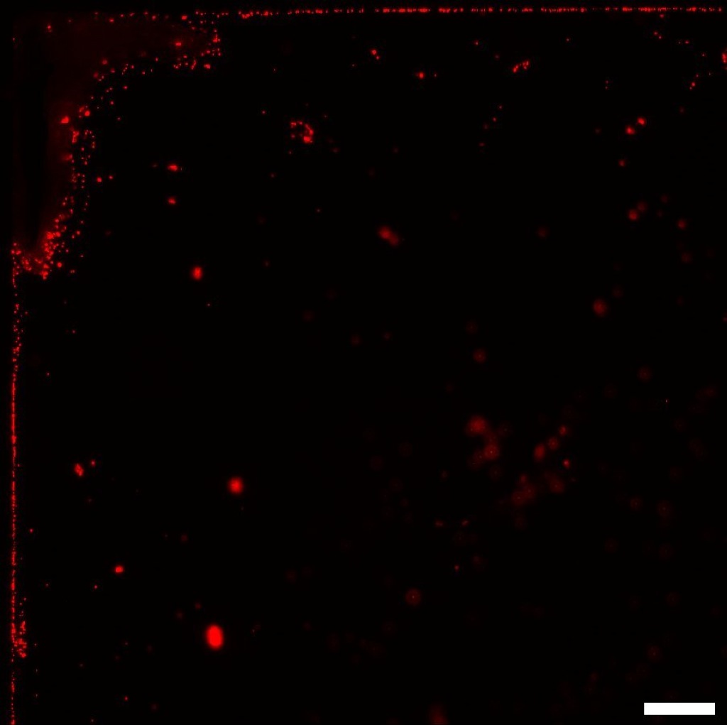
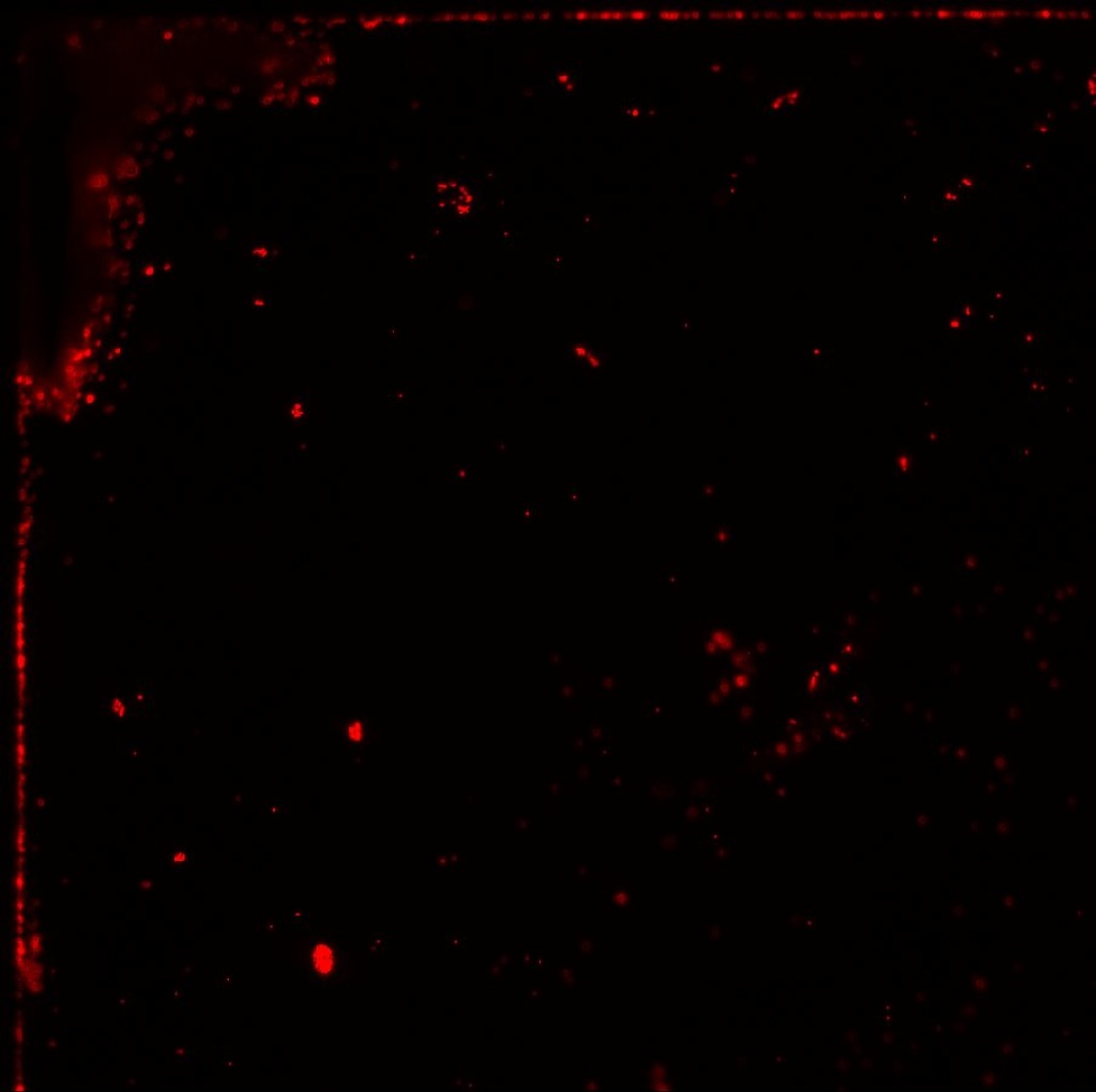
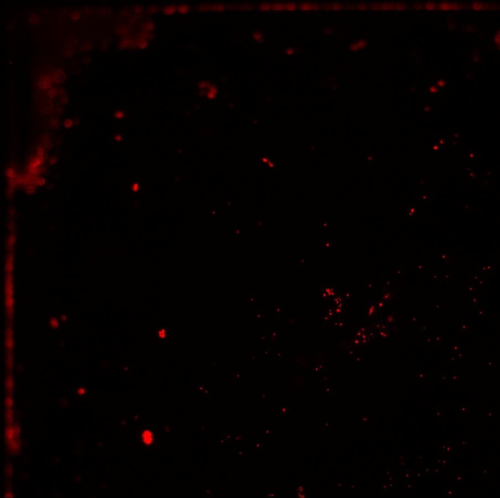
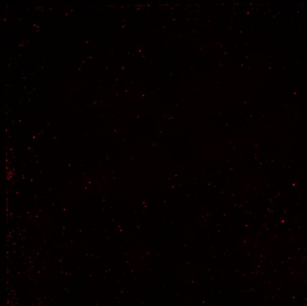
100W, Autoclaved

150W, Autoclaved

I also took a series of images on the 100W, Not Autoclaved membrane where the first is in focus, second is 10um above that focus, and third is 10um below the original focus.

It does seem most beads remain in the same focal plane and blur equally between the images, implying a lack of sagging.
Images have been taken such to repeat the experiment in the first post regarding the fluorescent intensity of the beads through the membrane, and I will update that as soon as possible as well.
Great news.
These samples were fabricated using new data that suggests a longer (8 hour) anneal time in forming gas may keep the SiO2 stress from “drifting” over time. Previously I was performing a 1 hour anneal on this material.
JP
So we changed two variables: thickness and anneal time. Which of the two variables reduced the sags?
The data also shows that there is not a significant difference in film stress over time when comparing 100nm and 300nm-thick PECVD SiO2. In fact, the stress for the two film thickness are virtually identical over 3 weeks.
I assume that most of the stress data has been taken with wafers that have not been etched, correct? Do we also have membranes that have not wrinkled for over 3 weeks? The exposure to the etch and formation of a membrane is different from a film sitting on a wafer, so hopefully the stability testing of membranes is also ongoing and they are staying flat.
Regarding these sag tests, is there liquid on both sides of the membrane during all tests? Do you think that the fact that the most bowing was seen on the non-porous membrane is a factor? Naively, I would expect less bow in a porous membrane that intrinsically equalizes pressure on both sides….
Thanks!
To answer the question about the sag tests, liquid is placed on both sides of the membrane via standard CytoVu protocol with use of top & bottom ‘gaskets’. It is possible that the more porous membranes contributed to some amount of stabilization, but even just to the eye the nonporous membranes have clear wrinkles. I would be led to believe that bowing caused by such wouldn’t be mitigated by pressure equalizatoin entirely, since the wrinkles would need to distribute somehow.
Thanks Zach! I agree that you if you start wrinkled you will have more of something, although I would not call it “sag” as defined above. If the wrinkled membranes were porous, you would probably see a similar “wrinkle” to what you see dry. I assume this would still be a problem? Say you have wrinkle of 5-10um instead of a low-pressure sag of 40um? Maybe this would not be an issue?
To Jim’s point, instead of changing 2 things, we have actually changed 3 or more variables as we compare across these samples in terms of porous/nonporous. My only real concern here is whether the new porous glass membranes will have any degradation in performance if they wrinkle up after some time on the shelf or after exposure to an odd buffer or chemistry. This material is not a fully stable SiO2, and I’m sure it would wrinkle up after a high temperature exposure (600C or higher) or certain chemical treatments. I’m hoping that no “normal” biological use will wrinkle them, but it would still be useful to understand how a wrinkled version of these new porous membranes would behave.
If you are interested in a direct comparison of these membranes in wrinkled form, they would just need to be annealed at 800-900C in O2, probably, or just repeat the 1000C anneal that your non-porous samples saw….
Thanks!
Thanks for the insight/input! I do think that the wrinkles without sagging could be problematic, as cells would see that and may be geometrically influenced (even if not detrimental to the cells, could still be an uncontrolled factor if looking at angiogenesis or other behaviors). I’m not sure which would be more of an issue, as I haven’t performed much cell culture on the highly wrinkled membranes.
Thank you. It sounds like we need to be absolutely certain that the new porous SiO2 materials don’t wrinkled under any reasonable circumstances.
Are any of these tests ongoing? I don’t know what cell bio people consider “normal” conditions, but maybe soaking for a week in X, Y, Z buffers representing extremes pH or other chemical environment? It sounds like they stay flat through autoclaving, which is a good start, but we should probably test over several autoclaving cycles and various storage conditions and time as well….
I haven’t had any ongoing tests in this, no, but I can check with Dr. Gaborski if we would want to maybe subject them to some time in cell culture media or some other condition which would be indicative of normal cell culture use.
I recommend we do the following tests. Very acidic or basic buffers would not be appropriate for cell culture, but let’s try some sodium bicarbonate since we’ve seen membrane dissolution in basic conditions previously:
1. Most typical cell culture condition – pH should be 7.4-7.6 in the incubator. DMEM w/ 10% FBS and antibiotics for 1 week in the incubator (no cells). After 1 week, suspend fluorescent beads as previously done and image multiple focal planes. After imaging, rinse with ddH20 and inspect for wrinkling.
2. Slightly basic condition, the pH should be ~8.2.
0.1M Sodium Bicarbonate 1 week in the incubator (no cells, FBS or antibiotics). After 1 week, suspend fluorescent beads as previously done and image multiple focal planes. After imaging, rinse with ddH20 and inspect for wrinkling.
3. Standard salt buffer in non-incubator conditions, the pH should be 7.4.
1X PBS for 1 week on the benchtop (no cells, FBS or antibiotics). After 1 week, suspend fluorescent beads as previously done and image multiple focal planes. After imaging, rinse with ddH20 and inspect for wrinkling.
Please do this for both the 100W and 150W deposition conditions. Use whichever porosity we have more of in stock.